Mri spine anatomy radiology pdf
Mri spine anatomy radiology pdf
1 Medford Radiological Group, 692 Murphy Rd., Medford, OR 97504. 2 Department of Radiology, Baylor College of Medicine, One Baylor Plaza, Houston, TX 77030. 3 Department of Pediatric Radiology, F3503, University of Michigan, 1500 E. Medical Center Dr., …
CT and MRI Sequences Viewable as Presented in PACS 1, 2 (PACS = Picture Archiving and Communication System, the commonly used method for electronic storage and viewing of radiologic images) Spine Images
Atlas of MRI Anatomy of the Abdomen. This photo gallery presents the anatomy of the abdomen by means of MRI. Axial T1-weighted images Axial T1-weighted fat …
MATERIALS AND METHODS: Adult patients undergoing cervical spine MR imaging with both T2-SPACE and T2-FSE sequences for radiculopathy or myelopathy between September 2014 and February 2015 were included.
Cornelius von Morze Ph.D. is a Senior Bioengineer scientist in the Department of Radiology & Biomedical Imaging at UCSF with extensive experience in preclinical applications for imaging hyperpolarized carbon-13 contrast agents for investigations of metabolism, perfusion, and transport. He is also developing a new research program focused on renal HP MRI studies.
Musculoskeletal imaging is a subspecialty of diagnostic radiology which involves ordering and interpreting medical images of bones, joints and associated soft …
Basic radiological anatomy of the brain and spine with annotated CT and MRI images covering the brain, including the brainstem structures and ventricles, and whole spine.
Lectures . Most of the lectures covering clinical topics contain radiological images. CXR Clinicopathological Correlation ; A.J. Chandrasekhar, MD
22/07/2016 · Magnetic resonance imaging (MRI) has been playing an increasingly important role in the spinal trauma patients due to high sensitivity for detection of acute soft tissue and cord injuries.
Spine Anatomy Neuroradiology – Google Sites

HeadNeckBrainSpine
INTRODUCTION. Magnetic resonance imaging (MRI) has become the examination of choice for imaging the spine and its contents. Although diseases of the spine are very common, clinical syndromes may mimic each other, necessitating imaging such as MRI …
The MRI protocol for examination of the lumbar spine in patients with symptoms of nerve compression is quite simple. Basically we rely on the sagittal T1W- and T2W-images and correlate the findings with the transverse T2W-images of the levels of suspected pathology.
An MRI examination of the spine shows the anatomy of the vertebrae that make up the spine, ligaments that hold the vertebrae together, as well as the disks, spinal cord and the spaces between the vertebrae through which nerves pass.
An MRI of the spine shows the anatomy of the vertebrae that make up the spine, as well as the disks, spinal cord and the spaces between the vertebrae through which nerves pass. Currently, MRI is the most sensitive imaging test of the spine in routine clinical practice.
The Thoraco-Lumbar Injury Classification and Severity score (TLICS) is a classification system for thoracolumbar spine injuries, designed to assist in clinical management. Unlike other classifications, the TLICS is an easy scoring system that depicts the features important in predicting spinal
MRI of Lumbar Spine Date: May 1, 2007 Updated: April 28, 2009 Exeeccuuttiivvee SSuu mmmaarryy Purpose: To identify and discourage the inappropriate use of high tech, high cost diagnostic imaging. Why was this Issue Selected: The indiscriminate use of expensive imaging exams for common and uncomplicated clinical presentations of the back and spine, e.g. low back pain, have contributed to …
Neuroradiology: the Essentials with MR and CT is written both to be read from cover to cover and to be used as a quick reference in the midst of a busy clinical day. Designed as a practical educational resource for clinical Neuroradiology, the text is divided into three sections – the brain, head and neck, and spine. Care has been taken for the text to be inclusive, yet focusing on commonly
MR Imaging of the Spine and Spinal Cord Pdf Magnetic resonance imaging has become an increasingly beneficial tool for the radiologic evaluation of complex spine diseases. However, due to the many variables implicit in MR imaging technique, considerable experience and expertise are necessary to diagnose with confidence.

This MRI cervical spine (C Spine) cross sectional anatomy tool is absolutely free to use. Use the mouse scroll wheel to move the images up and down alternatively use the tiny arrows (>>) on both side of the image to move the images.
Diffusion MRI is an active area of research that produces an unprecedented wealth of information on white matter anatomy. Future refinement of diffusion MRI tools for mapping white matter pathways will continue to advance tissue characterization, surgical planning, and follow-up of brain tumor patients.
11/09/2015 · All the anatomy you need to understand for reading a Lumbar Spine MRI.
radiology to form pictures of the anatomy and the physiological processes of the body in both health and disease. MRI scanners use strong magnetic fields, magnetic field gradients, and radio waves to generate images of the organs in the body. MRI does not involve X-rays or the use of ionizing radiation, which distinguishes Thu, 13 Dec 2018 01:27:00 GMT Magnetic resonance imaging
Clinical considerations are particularly important in the context of Cervical spine (C-spine) injury. This is because normal C-spine X-rays cannot exclude significant injury, and because a missed C-spine fracture can lead to death, or life long neurological deficit.
Download imaging anatomy of the human spine or read online here in PDF or EPUB. Please click button to get imaging anatomy of the human spine book now. All books are in clear copy here, and all files are secure so don’t worry about it.
Some spine examinations may require an injection of an MRI contrast (gadolinium) to help better visualise anatomy. If this is required, it will be discussed with you before proceeding. If this is required, it will be discussed with you before proceeding.
SPINE IMAGING GUIDELINES SP-1~GENERAL GUIDELINES Procedure Codes Associated with Spine Imaging MRI/MRA CPT® Cervical MRI without contrast 72141
Spinal anatomy Radiology Reference Article Radiopaedia.org
ONLINE MRI & CT SECTIONAL ANATOMY Kenneth K. F. Ho Bachelor of Medicine, Bachelor of Surgery (University of Hong Kong) Fellow, Hong Kong College of Radiologists Fellow, Hong Kong Academy of Medicine (Radiology)
Magnetic Resonance Imaging (MRI): Lumbar Spine. What It Is. Magnetic resonance imaging (MRI) of the lumbar spine is a safe and painless test that uses a magnetic field and radio waves to produce detailed pictures of the lumbar spine (the bones, disks, and other structures in the lower back).
25/01/2012 · Dr. Mamdouh Mahfouz Radiologic Anatomy of spine Spine Imaging Series SSR Radiology Diploma Cairo University, School of Medicine Kasr AlAiny School of Medicine Egypt.
A normal disc is composed of a central nucleus pulposus and peripheral annulus fibrosus. The disc is within the boundaries of the disc space, as defined, craniad and caudad by the vertebral body end plates and peripherally by the planes of the outer edges of the vertebral apophyses.
9.1 Radiographic anatomy of the spine 187 9.2 Spine trauma 188 9.3 Neck pain 195 9.4 Low back pain 196 9.5 Specific back pain syndromes 198 9.6 Sciatica 203 9 Spine 9.1 RADIOGRAPHIC ANATOMY OF THE SPINE Anatomical features of each vertebral body that can be identified radiographically (Figs 9.1, 9.2 and 9.3) include: • Anterior vertebral body • Posterior arch formed by pedicles and lamina – rehabilitation of the spine a practitioners manual pdf download MRI Anatomy and Positioning Series Module 8: Cardiac Imaging 4 Introduction Welcome to the Hitachi Medical Systems America, Inc. MRI Anatomy and Positioning Series.
Although the ovine spine is a useful research model for intervertebral disc pathology and vertebral surgery, there is little peer-reviewed information regarding the MRI anatomy of the ovine spine.
Sectional Anatomy by MRI and CT, 4th Edition Imaging Anatomy: Musculoskeletal, 2nd Edition Succeeding in the FRCR Part 1 Anatomy Exam : An Illustrated Guide Including 20 Mock Examinations Comprising 400 Images
Welcome to HeadNeckBrainSpine, a website intended for those interested in neuroradiology anatomy and learning from neuroradiology cases. To navigate the …
Background. With improved accessibility and increasing use of magnetic resonance imaging (MRI) to evaluate low back pain, general practitioners are exposed to a set of recommended terminology used among the various specialties involved in lumbar spinal conditions.
Anatomy of the lumbar vertebrae (cross-sectional imaging on T1, T2 and 3D MR) Lumbar spine anatomy on MRI (Magnetic Resonance Imaging) Anatomy of the lumbar spine using cross-sectional imaging (MR T1 and T2 weighted; sagittal, coronal and transverse slices)
To help understand how to plot slices please print out the “Pulse Radiology MR Clinical Notes”, draw your corresponding anatomy and how to plot slices for each exam.
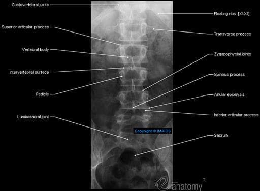
Radiology Anatomy of the Spine and Upper Extremity Timothy J. Mosher, MD Department of Radiology Phone: 531-4566 Email: tmosher@psu.edu. Learning Objective • Identify anatomic structures of the spine and upper extremities on standard radiographic and cross-sectional images. SPINE. Visual Human Sagittal View 7 Cervical Vertebra 12 Thoracic Vertebra 5 Lumbar Vertebra 5 Sacral Vertebra …
MRI Anatomy and Positioning Series Module 7: Neuro Imaging 5 . Introduction . Welcome to the Hitachi Medical Systems America, Inc. MRI Anatomy and Positioning Series. We offer teaching modules to allow users of Hitachi MRI scanners to review anatomy that will be seen on various MRI exams, and to enhance their positioning skills. Competent positioningensures the best possible …
This MRI cervical spine sagittal cross sectional anatomy tool is absolutely free to use. Use the mouse scroll wheel to move the images up and down alternatively use the tiny arrows (>>)on both side of the image to move the images.
Homepage – Welcome to w-radiology.com This web site is dedicated to the radiology and it is designed to work perfectly on desktop, laptop, and mobile devices (responsive design). Warning: Redesign of programming and content of this website during April 2015.
Magnetic Resonance Imaging (MRI) is a diagnostic imaging modality which uses radio waves and magnetic field to image the human body by aligning the magnetic nuclei (protons) of the body in a
Lumbar spine anatomy on MRI (Magnetic Resonance Imaging)
12/12/2013 · Keywords: Cervical spine, Trauma, Fracture, Computed tomography, Magnetic resonance imaging MDCT and MRI In most emergency departments, MDCT is the fastest and most practical study for cervical spine injury following trauma.
Teaching radiological anatomy 75 Fig. 3. Conventional Radiology. (A) Anteroposterior radiograph of abdomen, (B) anteroposterior radiograph of hand, (C) elimination urog-
Sonography of the neonatal spine is now accepted as a highly sensitive, readily available screening study that can be used to evaluate various anomalies of the lumbar spine …
An MRI examination of the spine shows the anatomy of the vertebrae that make up the spine, ligaments that hold the vertebrae together, as well as the disks, spinal cord and …
MR Imaging of the Spine and Spinal Cord Free Pdf Download

Sonography of the Neonatal Spine Part 1 Normal Anatomy
In part 1 of this pictorial essay, we discuss lumbar spine embryology, sonography techniques and indications, normal anatomy, and developmental variations and pitfalls that may simulate disease. Part 2 covers abnormal entities.
Spinal anatomy encompasses the anatomy of all osseous and soft tissue structures of the spine, the spinal cord and its supporting structures. This anatomy section promotes the use of the Terminologia Anatomica , the global standard for correct gross anatomical nomenclature.
Download Atlas of Human Anatomy on MRI: Spine Extremities Joints PDF. Hariqbal singh md dmrd professor & head, department of radiology parvez sheik mbbs dmre consultant radiology, department of radiology both at shrimati kashibai navale medical college, pune, maharashtra, india
To discuss routine Spine MRI protocols, and the sequences’ importance in an exam To identify the “H” sign of gray matter with the spinal cord, axial section To discuss conus medullaris vs. cauda equine, ending in the filum terminale
What It Is. Magnetic resonance imaging (MRI) of the lumbar spine is a safe and painless test that uses a magnetic field and radio waves to produce detailed pictures of the lumbar spine (the bones, disks, and other structures in the lower back).
Spine Radiology x ray ct mri normal anatomy – Free ebook download as Powerpoint Presentation (.ppt / .pptx), PDF File (.pdf), Text File (.txt) or view presentation slides online.
Spine MRI Radiology Key

Radiology basics Head anatomy
Emergency radiologic evaluation of the pediatric cervical spine can be challenging because of the confusing appearance of synchondroses, normal anatomic variants, and injuries that are unique to children. Cervical spine injuries in children are usually seen in the upper cervical region owing to the unique biomechanics and anatomy of the
Normal MR Imaging Anatomy of the Knee Saifuddin Vohra, DO, George Arnold, MD, Shashin Doshi, MD, David Marcantonio, MD* There are several keys to successfully interpreting
Discover (and save!) your own Pins on Pinterest. Color MRI Image Lumbar Spine Anatomy . Color MRI Image Lumbar Spine Anatomy. Visit. Discover ideas about Nerve Anatomy. What Is a Lumbar MRI? An MRI uses magnets and radio waves to capture images inside your body without making a surgical incision. The scan allows your doctor to. Nerve Anatomy Spinal Cord Neck Pain Radiology …
DOWNLOAD PDF MRI AND CT OF THE FEMALE PELVIS (MEDICAL RADIOLOGY DIAGNOSTIC IMAGING) Chapter 1 : Imaging the female pelvis: When should MRI be considered? MRI and CT exquisitely depict the anatomy of the female pelvis and offer fascinating diagnostic possibilities in women with pelvic disorders. This volume provides a comprehensive account of the use of these cross-sectional imaging …
The lumbar spine consists of five adjacent vertebrae of the lower vertebral column. They participate in the lumbar lordosis, a natural curve in the spine, that is convex anteriorly. Articulations of the facet (zygapophyseal) joints permit flexi…
Imaging Anatomy of the Human Spine A Comprehensive Atlas Including Adjacent Structures Scott E. Forseen, MD Assistant Professor, Neuroradiology Section Department of Radiology and Imaging Georgia Regents University Augusta, Georgia Neil M. Borden, MD Neuroradiologist Associate Professor of Radiology The University of Vermont Medical Center Burlington, Vermont. …
Segmental Anatomy. X-Ray, CT and MRI. MRI. Miscellaneous. Cervical Spine. Dens. Spinal Cord. Foramen Transversarium. Lines. Anterior Vertebral line. Posterior
Magnetic Resonance Imaging (MRI) Spine


Color MRI Image Lumbar Spine Anatomy Mri Scans Pinterest
https://simple.wikipedia.org/wiki/Spine
Lumbar spine Radiology Reference Article Radiopaedia.org
– MDCT and MRI evaluation of cervical spine trauma
Magnetic Resonance Imaging (MRI) Spine – RadiologyInfo.org


The Radiology Assistant Spine – Lumbar Disc Herniation
Mri In Epilepsy Medical Radiology
Spine Lumbar Disc Nomenclature 2.0 – Radiology Assistant
Magnetic Resonance Imaging (MRI) Lumbar Spine (for Parents)
Homepage – Welcome to w-radiology.com This web site is dedicated to the radiology and it is designed to work perfectly on desktop, laptop, and mobile devices (responsive design). Warning: Redesign of programming and content of this website during April 2015.
Musculoskeletal imaging is a subspecialty of diagnostic radiology which involves ordering and interpreting medical images of bones, joints and associated soft …
The lumbar spine consists of five adjacent vertebrae of the lower vertebral column. They participate in the lumbar lordosis, a natural curve in the spine, that is convex anteriorly. Articulations of the facet (zygapophyseal) joints permit flexi…
MR Imaging of the Spine and Spinal Cord Pdf Magnetic resonance imaging has become an increasingly beneficial tool for the radiologic evaluation of complex spine diseases. However, due to the many variables implicit in MR imaging technique, considerable experience and expertise are necessary to diagnose with confidence.
CT and MRI Sequences Viewable as Presented in PACS 1, 2 (PACS = Picture Archiving and Communication System, the commonly used method for electronic storage and viewing of radiologic images) Spine Images
A normal disc is composed of a central nucleus pulposus and peripheral annulus fibrosus. The disc is within the boundaries of the disc space, as defined, craniad and caudad by the vertebral body end plates and peripherally by the planes of the outer edges of the vertebral apophyses.
Lectures . Most of the lectures covering clinical topics contain radiological images. CXR Clinicopathological Correlation ; A.J. Chandrasekhar, MD
MATERIALS AND METHODS: Adult patients undergoing cervical spine MR imaging with both T2-SPACE and T2-FSE sequences for radiculopathy or myelopathy between September 2014 and February 2015 were included.
22/07/2016 · Magnetic resonance imaging (MRI) has been playing an increasingly important role in the spinal trauma patients due to high sensitivity for detection of acute soft tissue and cord injuries.
Cervical or Thoracic Spine MRI Sound Radiology
Radiology Anatomy of the Spine and Upper Extremity
Clinical considerations are particularly important in the context of Cervical spine (C-spine) injury. This is because normal C-spine X-rays cannot exclude significant injury, and because a missed C-spine fracture can lead to death, or life long neurological deficit.
This MRI cervical spine sagittal cross sectional anatomy tool is absolutely free to use. Use the mouse scroll wheel to move the images up and down alternatively use the tiny arrows (>>)on both side of the image to move the images.
radiology to form pictures of the anatomy and the physiological processes of the body in both health and disease. MRI scanners use strong magnetic fields, magnetic field gradients, and radio waves to generate images of the organs in the body. MRI does not involve X-rays or the use of ionizing radiation, which distinguishes Thu, 13 Dec 2018 01:27:00 GMT Magnetic resonance imaging
MRI of Lumbar Spine Date: May 1, 2007 Updated: April 28, 2009 Exeeccuuttiivvee SSuu mmmaarryy Purpose: To identify and discourage the inappropriate use of high tech, high cost diagnostic imaging. Why was this Issue Selected: The indiscriminate use of expensive imaging exams for common and uncomplicated clinical presentations of the back and spine, e.g. low back pain, have contributed to …
Magnetic Resonance Imaging (MRI): Lumbar Spine. What It Is. Magnetic resonance imaging (MRI) of the lumbar spine is a safe and painless test that uses a magnetic field and radio waves to produce detailed pictures of the lumbar spine (the bones, disks, and other structures in the lower back).
Color MRI Image Lumbar Spine Anatomy Mri Scans Pinterest
The Radiology Assistant Spine – Lumbar Disc Herniation
An MRI examination of the spine shows the anatomy of the vertebrae that make up the spine, ligaments that hold the vertebrae together, as well as the disks, spinal cord and the spaces between the vertebrae through which nerves pass.
A normal disc is composed of a central nucleus pulposus and peripheral annulus fibrosus. The disc is within the boundaries of the disc space, as defined, craniad and caudad by the vertebral body end plates and peripherally by the planes of the outer edges of the vertebral apophyses.
The MRI protocol for examination of the lumbar spine in patients with symptoms of nerve compression is quite simple. Basically we rely on the sagittal T1W- and T2W-images and correlate the findings with the transverse T2W-images of the levels of suspected pathology.
Anatomy of the lumbar vertebrae (cross-sectional imaging on T1, T2 and 3D MR) Lumbar spine anatomy on MRI (Magnetic Resonance Imaging) Anatomy of the lumbar spine using cross-sectional imaging (MR T1 and T2 weighted; sagittal, coronal and transverse slices)
Normal MR Imaging Anatomy of the Knee Saifuddin Vohra, DO, George Arnold, MD, Shashin Doshi, MD, David Marcantonio, MD* There are several keys to successfully interpreting
Radiology Anatomy of the Spine and Upper Extremity Timothy J. Mosher, MD Department of Radiology Phone: 531-4566 Email: tmosher@psu.edu. Learning Objective • Identify anatomic structures of the spine and upper extremities on standard radiographic and cross-sectional images. SPINE. Visual Human Sagittal View 7 Cervical Vertebra 12 Thoracic Vertebra 5 Lumbar Vertebra 5 Sacral Vertebra …
Lectures . Most of the lectures covering clinical topics contain radiological images. CXR Clinicopathological Correlation ; A.J. Chandrasekhar, MD
22/07/2016 · Magnetic resonance imaging (MRI) has been playing an increasingly important role in the spinal trauma patients due to high sensitivity for detection of acute soft tissue and cord injuries.
radiology to form pictures of the anatomy and the physiological processes of the body in both health and disease. MRI scanners use strong magnetic fields, magnetic field gradients, and radio waves to generate images of the organs in the body. MRI does not involve X-rays or the use of ionizing radiation, which distinguishes Thu, 13 Dec 2018 01:27:00 GMT Magnetic resonance imaging
MR Imaging of the Spine and Spinal Cord Free Pdf Download
Mri In Epilepsy Medical Radiology
MR Imaging of the Spine and Spinal Cord Pdf Magnetic resonance imaging has become an increasingly beneficial tool for the radiologic evaluation of complex spine diseases. However, due to the many variables implicit in MR imaging technique, considerable experience and expertise are necessary to diagnose with confidence.
Spinal anatomy encompasses the anatomy of all osseous and soft tissue structures of the spine, the spinal cord and its supporting structures. This anatomy section promotes the use of the Terminologia Anatomica , the global standard for correct gross anatomical nomenclature.
A normal disc is composed of a central nucleus pulposus and peripheral annulus fibrosus. The disc is within the boundaries of the disc space, as defined, craniad and caudad by the vertebral body end plates and peripherally by the planes of the outer edges of the vertebral apophyses.
An MRI of the spine shows the anatomy of the vertebrae that make up the spine, as well as the disks, spinal cord and the spaces between the vertebrae through which nerves pass. Currently, MRI is the most sensitive imaging test of the spine in routine clinical practice.
Cervical or Thoracic Spine MRI Sound Radiology
Atlas of MRI Anatomy of the Abdomen w-radiology.com
Normal MR Imaging Anatomy of the Knee Saifuddin Vohra, DO, George Arnold, MD, Shashin Doshi, MD, David Marcantonio, MD* There are several keys to successfully interpreting
CT and MRI Sequences Viewable as Presented in PACS 1, 2 (PACS = Picture Archiving and Communication System, the commonly used method for electronic storage and viewing of radiologic images) Spine Images
11/09/2015 · All the anatomy you need to understand for reading a Lumbar Spine MRI.
Download imaging anatomy of the human spine or read online here in PDF or EPUB. Please click button to get imaging anatomy of the human spine book now. All books are in clear copy here, and all files are secure so don’t worry about it.
Diffusion MRI an overview ScienceDirect Topics
The Radiology Assistant Spine injury – TLICS Classification
Neuroradiology: the Essentials with MR and CT is written both to be read from cover to cover and to be used as a quick reference in the midst of a busy clinical day. Designed as a practical educational resource for clinical Neuroradiology, the text is divided into three sections – the brain, head and neck, and spine. Care has been taken for the text to be inclusive, yet focusing on commonly
Sectional Anatomy by MRI and CT, 4th Edition Imaging Anatomy: Musculoskeletal, 2nd Edition Succeeding in the FRCR Part 1 Anatomy Exam : An Illustrated Guide Including 20 Mock Examinations Comprising 400 Images
Welcome to HeadNeckBrainSpine, a website intended for those interested in neuroradiology anatomy and learning from neuroradiology cases. To navigate the …
Basic radiological anatomy of the brain and spine with annotated CT and MRI images covering the brain, including the brainstem structures and ventricles, and whole spine.
Radiology Anatomy of the Spine and Upper Extremity
The Radiology Assistant Spine injury – TLICS Classification
MRI Anatomy and Positioning Series Module 8: Cardiac Imaging 4 Introduction Welcome to the Hitachi Medical Systems America, Inc. MRI Anatomy and Positioning Series.
Lectures . Most of the lectures covering clinical topics contain radiological images. CXR Clinicopathological Correlation ; A.J. Chandrasekhar, MD
MRI of Lumbar Spine Date: May 1, 2007 Updated: April 28, 2009 Exeeccuuttiivvee SSuu mmmaarryy Purpose: To identify and discourage the inappropriate use of high tech, high cost diagnostic imaging. Why was this Issue Selected: The indiscriminate use of expensive imaging exams for common and uncomplicated clinical presentations of the back and spine, e.g. low back pain, have contributed to …
Anatomy of the lumbar vertebrae (cross-sectional imaging on T1, T2 and 3D MR) Lumbar spine anatomy on MRI (Magnetic Resonance Imaging) Anatomy of the lumbar spine using cross-sectional imaging (MR T1 and T2 weighted; sagittal, coronal and transverse slices)
MDCT and MRI evaluation of cervical spine trauma
Radiology Spine Imaging – Normal Anatomy – YouTube
Homepage – Welcome to w-radiology.com This web site is dedicated to the radiology and it is designed to work perfectly on desktop, laptop, and mobile devices (responsive design). Warning: Redesign of programming and content of this website during April 2015.
An MRI of the spine shows the anatomy of the vertebrae that make up the spine, as well as the disks, spinal cord and the spaces between the vertebrae through which nerves pass. Currently, MRI is the most sensitive imaging test of the spine in routine clinical practice.
Spinal anatomy encompasses the anatomy of all osseous and soft tissue structures of the spine, the spinal cord and its supporting structures. This anatomy section promotes the use of the Terminologia Anatomica , the global standard for correct gross anatomical nomenclature.
The Thoraco-Lumbar Injury Classification and Severity score (TLICS) is a classification system for thoracolumbar spine injuries, designed to assist in clinical management. Unlike other classifications, the TLICS is an easy scoring system that depicts the features important in predicting spinal
9.1 Radiographic anatomy of the spine 187 9.2 Spine trauma 188 9.3 Neck pain 195 9.4 Low back pain 196 9.5 Specific back pain syndromes 198 9.6 Sciatica 203 9 Spine 9.1 RADIOGRAPHIC ANATOMY OF THE SPINE Anatomical features of each vertebral body that can be identified radiographically (Figs 9.1, 9.2 and 9.3) include: • Anterior vertebral body • Posterior arch formed by pedicles and lamina
Cornelius von Morze Ph.D. is a Senior Bioengineer scientist in the Department of Radiology & Biomedical Imaging at UCSF with extensive experience in preclinical applications for imaging hyperpolarized carbon-13 contrast agents for investigations of metabolism, perfusion, and transport. He is also developing a new research program focused on renal HP MRI studies.
This MRI cervical spine (C Spine) cross sectional anatomy tool is absolutely free to use. Use the mouse scroll wheel to move the images up and down alternatively use the tiny arrows (>>) on both side of the image to move the images.
A normal disc is composed of a central nucleus pulposus and peripheral annulus fibrosus. The disc is within the boundaries of the disc space, as defined, craniad and caudad by the vertebral body end plates and peripherally by the planes of the outer edges of the vertebral apophyses.
SPINE IMAGING GUIDELINES SP-1~GENERAL GUIDELINES Procedure Codes Associated with Spine Imaging MRI/MRA CPT® Cervical MRI without contrast 72141
11/09/2015 · All the anatomy you need to understand for reading a Lumbar Spine MRI.
Emergency radiologic evaluation of the pediatric cervical spine can be challenging because of the confusing appearance of synchondroses, normal anatomic variants, and injuries that are unique to children. Cervical spine injuries in children are usually seen in the upper cervical region owing to the unique biomechanics and anatomy of the
Download Atlas of Human Anatomy on MRI: Spine Extremities Joints PDF. Hariqbal singh md dmrd professor & head, department of radiology parvez sheik mbbs dmre consultant radiology, department of radiology both at shrimati kashibai navale medical college, pune, maharashtra, india
INTRODUCTION. Magnetic resonance imaging (MRI) has become the examination of choice for imaging the spine and its contents. Although diseases of the spine are very common, clinical syndromes may mimic each other, necessitating imaging such as MRI …
Welcome to HeadNeckBrainSpine, a website intended for those interested in neuroradiology anatomy and learning from neuroradiology cases. To navigate the …
Home page w-radiology.com
Trauma X-ray Axial skeleton – Cervical spine – Normal
To help understand how to plot slices please print out the “Pulse Radiology MR Clinical Notes”, draw your corresponding anatomy and how to plot slices for each exam.
Although the ovine spine is a useful research model for intervertebral disc pathology and vertebral surgery, there is little peer-reviewed information regarding the MRI anatomy of the ovine spine.
1 Medford Radiological Group, 692 Murphy Rd., Medford, OR 97504. 2 Department of Radiology, Baylor College of Medicine, One Baylor Plaza, Houston, TX 77030. 3 Department of Pediatric Radiology, F3503, University of Michigan, 1500 E. Medical Center Dr., …
Musculoskeletal imaging is a subspecialty of diagnostic radiology which involves ordering and interpreting medical images of bones, joints and associated soft …
Cornelius von Morze Ph.D. is a Senior Bioengineer scientist in the Department of Radiology & Biomedical Imaging at UCSF with extensive experience in preclinical applications for imaging hyperpolarized carbon-13 contrast agents for investigations of metabolism, perfusion, and transport. He is also developing a new research program focused on renal HP MRI studies.
Imaging Anatomy of the Human Spine A Comprehensive Atlas Including Adjacent Structures Scott E. Forseen, MD Assistant Professor, Neuroradiology Section Department of Radiology and Imaging Georgia Regents University Augusta, Georgia Neil M. Borden, MD Neuroradiologist Associate Professor of Radiology The University of Vermont Medical Center Burlington, Vermont. …
Emergency radiologic evaluation of the pediatric cervical spine can be challenging because of the confusing appearance of synchondroses, normal anatomic variants, and injuries that are unique to children. Cervical spine injuries in children are usually seen in the upper cervical region owing to the unique biomechanics and anatomy of the
The Thoraco-Lumbar Injury Classification and Severity score (TLICS) is a classification system for thoracolumbar spine injuries, designed to assist in clinical management. Unlike other classifications, the TLICS is an easy scoring system that depicts the features important in predicting spinal
MRI Anatomy and Positioning Series Module 7: Neuro Imaging 5 . Introduction . Welcome to the Hitachi Medical Systems America, Inc. MRI Anatomy and Positioning Series. We offer teaching modules to allow users of Hitachi MRI scanners to review anatomy that will be seen on various MRI exams, and to enhance their positioning skills. Competent positioningensures the best possible …
Anatomy of the lumbar vertebrae (cross-sectional imaging on T1, T2 and 3D MR) Lumbar spine anatomy on MRI (Magnetic Resonance Imaging) Anatomy of the lumbar spine using cross-sectional imaging (MR T1 and T2 weighted; sagittal, coronal and transverse slices)
Segmental Anatomy. X-Ray, CT and MRI. MRI. Miscellaneous. Cervical Spine. Dens. Spinal Cord. Foramen Transversarium. Lines. Anterior Vertebral line. Posterior
Discover (and save!) your own Pins on Pinterest. Color MRI Image Lumbar Spine Anatomy . Color MRI Image Lumbar Spine Anatomy. Visit. Discover ideas about Nerve Anatomy. What Is a Lumbar MRI? An MRI uses magnets and radio waves to capture images inside your body without making a surgical incision. The scan allows your doctor to. Nerve Anatomy Spinal Cord Neck Pain Radiology …
Basic radiological anatomy of the brain and spine with annotated CT and MRI images covering the brain, including the brainstem structures and ventricles, and whole spine.
Spine Lumbar Disc Nomenclature 2.0 – Radiology Assistant
HeadNeckBrainSpine
Sectional Anatomy by MRI and CT, 4th Edition Imaging Anatomy: Musculoskeletal, 2nd Edition Succeeding in the FRCR Part 1 Anatomy Exam : An Illustrated Guide Including 20 Mock Examinations Comprising 400 Images
Atlas of MRI Anatomy of the Abdomen. This photo gallery presents the anatomy of the abdomen by means of MRI. Axial T1-weighted images Axial T1-weighted fat …
The Thoraco-Lumbar Injury Classification and Severity score (TLICS) is a classification system for thoracolumbar spine injuries, designed to assist in clinical management. Unlike other classifications, the TLICS is an easy scoring system that depicts the features important in predicting spinal
MRI Anatomy and Positioning Series Module 7: Neuro Imaging 5 . Introduction . Welcome to the Hitachi Medical Systems America, Inc. MRI Anatomy and Positioning Series. We offer teaching modules to allow users of Hitachi MRI scanners to review anatomy that will be seen on various MRI exams, and to enhance their positioning skills. Competent positioningensures the best possible …
Radiology Anatomy of the Spine and Upper Extremity Timothy J. Mosher, MD Department of Radiology Phone: 531-4566 Email: tmosher@psu.edu. Learning Objective • Identify anatomic structures of the spine and upper extremities on standard radiographic and cross-sectional images. SPINE. Visual Human Sagittal View 7 Cervical Vertebra 12 Thoracic Vertebra 5 Lumbar Vertebra 5 Sacral Vertebra …
To discuss routine Spine MRI protocols, and the sequences’ importance in an exam To identify the “H” sign of gray matter with the spinal cord, axial section To discuss conus medullaris vs. cauda equine, ending in the filum terminale
MRI Anatomy and Positioning Series Module 8: Cardiac Imaging 4 Introduction Welcome to the Hitachi Medical Systems America, Inc. MRI Anatomy and Positioning Series.
Musculoskeletal imaging is a subspecialty of diagnostic radiology which involves ordering and interpreting medical images of bones, joints and associated soft …
Background. With improved accessibility and increasing use of magnetic resonance imaging (MRI) to evaluate low back pain, general practitioners are exposed to a set of recommended terminology used among the various specialties involved in lumbar spinal conditions.
Lumbar Spine MRI Made Ridiculously Simple Anatomy YouTube
Sonography of the Neonatal Spine Part 1 Normal Anatomy
To discuss routine Spine MRI protocols, and the sequences’ importance in an exam To identify the “H” sign of gray matter with the spinal cord, axial section To discuss conus medullaris vs. cauda equine, ending in the filum terminale
This MRI cervical spine (C Spine) cross sectional anatomy tool is absolutely free to use. Use the mouse scroll wheel to move the images up and down alternatively use the tiny arrows (>>) on both side of the image to move the images.
Diffusion MRI is an active area of research that produces an unprecedented wealth of information on white matter anatomy. Future refinement of diffusion MRI tools for mapping white matter pathways will continue to advance tissue characterization, surgical planning, and follow-up of brain tumor patients.
CT and MRI Sequences Viewable as Presented in PACS 1, 2 (PACS = Picture Archiving and Communication System, the commonly used method for electronic storage and viewing of radiologic images) Spine Images
Discover (and save!) your own Pins on Pinterest. Color MRI Image Lumbar Spine Anatomy . Color MRI Image Lumbar Spine Anatomy. Visit. Discover ideas about Nerve Anatomy. What Is a Lumbar MRI? An MRI uses magnets and radio waves to capture images inside your body without making a surgical incision. The scan allows your doctor to. Nerve Anatomy Spinal Cord Neck Pain Radiology …
An MRI examination of the spine shows the anatomy of the vertebrae that make up the spine, ligaments that hold the vertebrae together, as well as the disks, spinal cord and …
Pediatric Cervical Spine Normal Anatomy Variants and
HeadNeckBrainSpine
Magnetic Resonance Imaging (MRI): Lumbar Spine. What It Is. Magnetic resonance imaging (MRI) of the lumbar spine is a safe and painless test that uses a magnetic field and radio waves to produce detailed pictures of the lumbar spine (the bones, disks, and other structures in the lower back).
Homepage – Welcome to w-radiology.com This web site is dedicated to the radiology and it is designed to work perfectly on desktop, laptop, and mobile devices (responsive design). Warning: Redesign of programming and content of this website during April 2015.
An MRI examination of the spine shows the anatomy of the vertebrae that make up the spine, ligaments that hold the vertebrae together, as well as the disks, spinal cord and …
Emergency radiologic evaluation of the pediatric cervical spine can be challenging because of the confusing appearance of synchondroses, normal anatomic variants, and injuries that are unique to children. Cervical spine injuries in children are usually seen in the upper cervical region owing to the unique biomechanics and anatomy of the
Welcome to HeadNeckBrainSpine, a website intended for those interested in neuroradiology anatomy and learning from neuroradiology cases. To navigate the …
Diffusion MRI is an active area of research that produces an unprecedented wealth of information on white matter anatomy. Future refinement of diffusion MRI tools for mapping white matter pathways will continue to advance tissue characterization, surgical planning, and follow-up of brain tumor patients.
Hyperpolarized Carbon-13 ! MRI Technology UCSF Radiology
Radiology Anatomy of the Spine and Upper Extremity
Clinical considerations are particularly important in the context of Cervical spine (C-spine) injury. This is because normal C-spine X-rays cannot exclude significant injury, and because a missed C-spine fracture can lead to death, or life long neurological deficit.
This MRI cervical spine sagittal cross sectional anatomy tool is absolutely free to use. Use the mouse scroll wheel to move the images up and down alternatively use the tiny arrows (>>)on both side of the image to move the images.
Homepage – Welcome to w-radiology.com This web site is dedicated to the radiology and it is designed to work perfectly on desktop, laptop, and mobile devices (responsive design). Warning: Redesign of programming and content of this website during April 2015.
Radiology Anatomy of the Spine and Upper Extremity Timothy J. Mosher, MD Department of Radiology Phone: 531-4566 Email: tmosher@psu.edu. Learning Objective • Identify anatomic structures of the spine and upper extremities on standard radiographic and cross-sectional images. SPINE. Visual Human Sagittal View 7 Cervical Vertebra 12 Thoracic Vertebra 5 Lumbar Vertebra 5 Sacral Vertebra …
Sonography of the neonatal spine is now accepted as a highly sensitive, readily available screening study that can be used to evaluate various anomalies of the lumbar spine …
Normal MR Imaging Anatomy of the Knee SmartView
Magnetic Resonance Imaging (MRI) Spine
The MRI protocol for examination of the lumbar spine in patients with symptoms of nerve compression is quite simple. Basically we rely on the sagittal T1W- and T2W-images and correlate the findings with the transverse T2W-images of the levels of suspected pathology.
Sectional Anatomy by MRI and CT, 4th Edition Imaging Anatomy: Musculoskeletal, 2nd Edition Succeeding in the FRCR Part 1 Anatomy Exam : An Illustrated Guide Including 20 Mock Examinations Comprising 400 Images
Teaching radiological anatomy 75 Fig. 3. Conventional Radiology. (A) Anteroposterior radiograph of abdomen, (B) anteroposterior radiograph of hand, (C) elimination urog-
radiology to form pictures of the anatomy and the physiological processes of the body in both health and disease. MRI scanners use strong magnetic fields, magnetic field gradients, and radio waves to generate images of the organs in the body. MRI does not involve X-rays or the use of ionizing radiation, which distinguishes Thu, 13 Dec 2018 01:27:00 GMT Magnetic resonance imaging
22/07/2016 · Magnetic resonance imaging (MRI) has been playing an increasingly important role in the spinal trauma patients due to high sensitivity for detection of acute soft tissue and cord injuries.
Segmental Anatomy. X-Ray, CT and MRI. MRI. Miscellaneous. Cervical Spine. Dens. Spinal Cord. Foramen Transversarium. Lines. Anterior Vertebral line. Posterior
Lumbar Spine MRI Made Ridiculously Simple Anatomy YouTube
Sonography of the Neonatal Spine Part 1 Normal Anatomy
MRI Anatomy and Positioning Series Module 8: Cardiac Imaging 4 Introduction Welcome to the Hitachi Medical Systems America, Inc. MRI Anatomy and Positioning Series.
An MRI of the spine shows the anatomy of the vertebrae that make up the spine, as well as the disks, spinal cord and the spaces between the vertebrae through which nerves pass. Currently, MRI is the most sensitive imaging test of the spine in routine clinical practice.
Magnetic Resonance Imaging (MRI) is a diagnostic imaging modality which uses radio waves and magnetic field to image the human body by aligning the magnetic nuclei (protons) of the body in a
Neuroradiology: the Essentials with MR and CT is written both to be read from cover to cover and to be used as a quick reference in the midst of a busy clinical day. Designed as a practical educational resource for clinical Neuroradiology, the text is divided into three sections – the brain, head and neck, and spine. Care has been taken for the text to be inclusive, yet focusing on commonly
CT and MRI Sequences Viewable as Presented in PACS 1, 2 (PACS = Picture Archiving and Communication System, the commonly used method for electronic storage and viewing of radiologic images) Spine Images
The Radiology Assistant Spine – Lumbar Disc Herniation
mri cervical spine sagittal anatomy Mrimaster.com
An MRI examination of the spine shows the anatomy of the vertebrae that make up the spine, ligaments that hold the vertebrae together, as well as the disks, spinal cord and the spaces between the vertebrae through which nerves pass.
9.1 Radiographic anatomy of the spine 187 9.2 Spine trauma 188 9.3 Neck pain 195 9.4 Low back pain 196 9.5 Specific back pain syndromes 198 9.6 Sciatica 203 9 Spine 9.1 RADIOGRAPHIC ANATOMY OF THE SPINE Anatomical features of each vertebral body that can be identified radiographically (Figs 9.1, 9.2 and 9.3) include: • Anterior vertebral body • Posterior arch formed by pedicles and lamina
INTRODUCTION. Magnetic resonance imaging (MRI) has become the examination of choice for imaging the spine and its contents. Although diseases of the spine are very common, clinical syndromes may mimic each other, necessitating imaging such as MRI …
Some spine examinations may require an injection of an MRI contrast (gadolinium) to help better visualise anatomy. If this is required, it will be discussed with you before proceeding. If this is required, it will be discussed with you before proceeding.
Imaging Anatomy of the Human Spine A Comprehensive Atlas Including Adjacent Structures Scott E. Forseen, MD Assistant Professor, Neuroradiology Section Department of Radiology and Imaging Georgia Regents University Augusta, Georgia Neil M. Borden, MD Neuroradiologist Associate Professor of Radiology The University of Vermont Medical Center Burlington, Vermont. …
To discuss routine Spine MRI protocols, and the sequences’ importance in an exam To identify the “H” sign of gray matter with the spinal cord, axial section To discuss conus medullaris vs. cauda equine, ending in the filum terminale
Anatomy of the lumbar vertebrae (cross-sectional imaging on T1, T2 and 3D MR) Lumbar spine anatomy on MRI (Magnetic Resonance Imaging) Anatomy of the lumbar spine using cross-sectional imaging (MR T1 and T2 weighted; sagittal, coronal and transverse slices)
Lectures . Most of the lectures covering clinical topics contain radiological images. CXR Clinicopathological Correlation ; A.J. Chandrasekhar, MD
MR Imaging of the Spine and Spinal Cord Free Pdf Download
Lumbar spine Radiology Reference Article Radiopaedia.org
Download imaging anatomy of the human spine or read online here in PDF or EPUB. Please click button to get imaging anatomy of the human spine book now. All books are in clear copy here, and all files are secure so don’t worry about it.
Basic radiological anatomy of the brain and spine with annotated CT and MRI images covering the brain, including the brainstem structures and ventricles, and whole spine.
An MRI of the spine shows the anatomy of the vertebrae that make up the spine, as well as the disks, spinal cord and the spaces between the vertebrae through which nerves pass. Currently, MRI is the most sensitive imaging test of the spine in routine clinical practice.
Although the ovine spine is a useful research model for intervertebral disc pathology and vertebral surgery, there is little peer-reviewed information regarding the MRI anatomy of the ovine spine.
The Thoraco-Lumbar Injury Classification and Severity score (TLICS) is a classification system for thoracolumbar spine injuries, designed to assist in clinical management. Unlike other classifications, the TLICS is an easy scoring system that depicts the features important in predicting spinal
MR Imaging of the Spine and Spinal Cord Pdf Magnetic resonance imaging has become an increasingly beneficial tool for the radiologic evaluation of complex spine diseases. However, due to the many variables implicit in MR imaging technique, considerable experience and expertise are necessary to diagnose with confidence.
In part 1 of this pictorial essay, we discuss lumbar spine embryology, sonography techniques and indications, normal anatomy, and developmental variations and pitfalls that may simulate disease. Part 2 covers abnormal entities.
CT and MRI Sequences Viewable as Presented in PACS 1, 2 (PACS = Picture Archiving and Communication System, the commonly used method for electronic storage and viewing of radiologic images) Spine Images
Discover (and save!) your own Pins on Pinterest. Color MRI Image Lumbar Spine Anatomy . Color MRI Image Lumbar Spine Anatomy. Visit. Discover ideas about Nerve Anatomy. What Is a Lumbar MRI? An MRI uses magnets and radio waves to capture images inside your body without making a surgical incision. The scan allows your doctor to. Nerve Anatomy Spinal Cord Neck Pain Radiology …
MATERIALS AND METHODS: Adult patients undergoing cervical spine MR imaging with both T2-SPACE and T2-FSE sequences for radiculopathy or myelopathy between September 2014 and February 2015 were included.
Trauma X-ray Axial skeleton – Cervical spine – Normal
Pediatric Cervical Spine Normal Anatomy Variants and
Atlas of MRI Anatomy of the Abdomen. This photo gallery presents the anatomy of the abdomen by means of MRI. Axial T1-weighted images Axial T1-weighted fat …
An MRI examination of the spine shows the anatomy of the vertebrae that make up the spine, ligaments that hold the vertebrae together, as well as the disks, spinal cord and …
Cornelius von Morze Ph.D. is a Senior Bioengineer scientist in the Department of Radiology & Biomedical Imaging at UCSF with extensive experience in preclinical applications for imaging hyperpolarized carbon-13 contrast agents for investigations of metabolism, perfusion, and transport. He is also developing a new research program focused on renal HP MRI studies.
In part 1 of this pictorial essay, we discuss lumbar spine embryology, sonography techniques and indications, normal anatomy, and developmental variations and pitfalls that may simulate disease. Part 2 covers abnormal entities.
Spinal anatomy encompasses the anatomy of all osseous and soft tissue structures of the spine, the spinal cord and its supporting structures. This anatomy section promotes the use of the Terminologia Anatomica , the global standard for correct gross anatomical nomenclature.
Magnetic Resonance Imaging (MRI) is a diagnostic imaging modality which uses radio waves and magnetic field to image the human body by aligning the magnetic nuclei (protons) of the body in a
Diffusion MRI is an active area of research that produces an unprecedented wealth of information on white matter anatomy. Future refinement of diffusion MRI tools for mapping white matter pathways will continue to advance tissue characterization, surgical planning, and follow-up of brain tumor patients.
An MRI of the spine shows the anatomy of the vertebrae that make up the spine, as well as the disks, spinal cord and the spaces between the vertebrae through which nerves pass. Currently, MRI is the most sensitive imaging test of the spine in routine clinical practice.
MRI Anatomy and Positioning Series Module 8: Cardiac Imaging 4 Introduction Welcome to the Hitachi Medical Systems America, Inc. MRI Anatomy and Positioning Series.
radiology to form pictures of the anatomy and the physiological processes of the body in both health and disease. MRI scanners use strong magnetic fields, magnetic field gradients, and radio waves to generate images of the organs in the body. MRI does not involve X-rays or the use of ionizing radiation, which distinguishes Thu, 13 Dec 2018 01:27:00 GMT Magnetic resonance imaging
MATERIALS AND METHODS: Adult patients undergoing cervical spine MR imaging with both T2-SPACE and T2-FSE sequences for radiculopathy or myelopathy between September 2014 and February 2015 were included.
Download Atlas of Human Anatomy on MRI: Spine Extremities Joints PDF. Hariqbal singh md dmrd professor & head, department of radiology parvez sheik mbbs dmre consultant radiology, department of radiology both at shrimati kashibai navale medical college, pune, maharashtra, india
Segmental Anatomy. X-Ray, CT and MRI. MRI. Miscellaneous. Cervical Spine. Dens. Spinal Cord. Foramen Transversarium. Lines. Anterior Vertebral line. Posterior
Normal MR Imaging Anatomy of the Knee SmartView
MRI spine anatomy free MRI axial cervical spine anatomy
To discuss routine Spine MRI protocols, and the sequences’ importance in an exam To identify the “H” sign of gray matter with the spinal cord, axial section To discuss conus medullaris vs. cauda equine, ending in the filum terminale
9.1 Radiographic anatomy of the spine 187 9.2 Spine trauma 188 9.3 Neck pain 195 9.4 Low back pain 196 9.5 Specific back pain syndromes 198 9.6 Sciatica 203 9 Spine 9.1 RADIOGRAPHIC ANATOMY OF THE SPINE Anatomical features of each vertebral body that can be identified radiographically (Figs 9.1, 9.2 and 9.3) include: • Anterior vertebral body • Posterior arch formed by pedicles and lamina
11/09/2015 · All the anatomy you need to understand for reading a Lumbar Spine MRI.
25/01/2012 · Dr. Mamdouh Mahfouz Radiologic Anatomy of spine Spine Imaging Series SSR Radiology Diploma Cairo University, School of Medicine Kasr AlAiny School of Medicine Egypt.
An MRI of the spine shows the anatomy of the vertebrae that make up the spine, as well as the disks, spinal cord and the spaces between the vertebrae through which nerves pass. Currently, MRI is the most sensitive imaging test of the spine in routine clinical practice.
Cornelius von Morze Ph.D. is a Senior Bioengineer scientist in the Department of Radiology & Biomedical Imaging at UCSF with extensive experience in preclinical applications for imaging hyperpolarized carbon-13 contrast agents for investigations of metabolism, perfusion, and transport. He is also developing a new research program focused on renal HP MRI studies.
Sectional Anatomy by MRI and CT, 4th Edition Imaging Anatomy: Musculoskeletal, 2nd Edition Succeeding in the FRCR Part 1 Anatomy Exam : An Illustrated Guide Including 20 Mock Examinations Comprising 400 Images
Diffusion MRI is an active area of research that produces an unprecedented wealth of information on white matter anatomy. Future refinement of diffusion MRI tools for mapping white matter pathways will continue to advance tissue characterization, surgical planning, and follow-up of brain tumor patients.
A normal disc is composed of a central nucleus pulposus and peripheral annulus fibrosus. The disc is within the boundaries of the disc space, as defined, craniad and caudad by the vertebral body end plates and peripherally by the planes of the outer edges of the vertebral apophyses.
In part 1 of this pictorial essay, we discuss lumbar spine embryology, sonography techniques and indications, normal anatomy, and developmental variations and pitfalls that may simulate disease. Part 2 covers abnormal entities.
Radiologic Anatomy Self Test Images of Normal Structure
Imaging Anatomy of the Human Spine A Comprehensive Atlas Including Adjacent Structures Scott E. Forseen, MD Assistant Professor, Neuroradiology Section Department of Radiology and Imaging Georgia Regents University Augusta, Georgia Neil M. Borden, MD Neuroradiologist Associate Professor of Radiology The University of Vermont Medical Center Burlington, Vermont. …
Sonography of the Neonatal Spine Part 1 Normal Anatomy
Download imaging anatomy of the human spine or read online here in PDF or EPUB. Please click button to get imaging anatomy of the human spine book now. All books are in clear copy here, and all files are secure so don’t worry about it.
Radiology Anatomy of the Spine and Upper Extremity
MRI Lumbar Spine Missouri Department of Social Services
25/01/2012 · Dr. Mamdouh Mahfouz Radiologic Anatomy of spine Spine Imaging Series SSR Radiology Diploma Cairo University, School of Medicine Kasr AlAiny School of Medicine Egypt.
Radiology basics Head anatomy
Cervical or Thoracic Spine MRI Sound Radiology
In part 1 of this pictorial essay, we discuss lumbar spine embryology, sonography techniques and indications, normal anatomy, and developmental variations and pitfalls that may simulate disease. Part 2 covers abnormal entities.
MR Imaging of the Spine and Spinal Cord Free Pdf Download
Musculoskeletal Imaging InsideRadiology
Magnetic Resonance Imaging (MRI) Spine – RadiologyInfo.org
An MRI of the spine shows the anatomy of the vertebrae that make up the spine, as well as the disks, spinal cord and the spaces between the vertebrae through which nerves pass. Currently, MRI is the most sensitive imaging test of the spine in routine clinical practice.
Musculoskeletal Imaging InsideRadiology
ONLINE MRI & CT SECTIONAL ANATOMY Kenneth K. F. Ho Bachelor of Medicine, Bachelor of Surgery (University of Hong Kong) Fellow, Hong Kong College of Radiologists Fellow, Hong Kong Academy of Medicine (Radiology)
Diffusion MRI an overview ScienceDirect Topics
Sonography of the Neonatal Spine Part 1 Normal Anatomy
Magnetic Resonance Imaging (MRI) Anatomy of the Ovine
The Thoraco-Lumbar Injury Classification and Severity score (TLICS) is a classification system for thoracolumbar spine injuries, designed to assist in clinical management. Unlike other classifications, the TLICS is an easy scoring system that depicts the features important in predicting spinal
Sonography of the Neonatal Spine Part 1 Normal Anatomy
Spine Lumbar Disc Nomenclature 2.0 – Radiology Assistant
Lumbar Spine MRI Made Ridiculously Simple Anatomy YouTube
1 Medford Radiological Group, 692 Murphy Rd., Medford, OR 97504. 2 Department of Radiology, Baylor College of Medicine, One Baylor Plaza, Houston, TX 77030. 3 Department of Pediatric Radiology, F3503, University of Michigan, 1500 E. Medical Center Dr., …
mri cervical spine sagittal anatomy Mrimaster.com
Diffusion MRI an overview ScienceDirect Topics
Atlas of MRI Anatomy of the Abdomen. This photo gallery presents the anatomy of the abdomen by means of MRI. Axial T1-weighted images Axial T1-weighted fat …
MR Imaging of the Spine and Spinal Cord Free Pdf Download
MRI Anatomy and Positioning Series Module 8: Cardiac Imaging 4 Introduction Welcome to the Hitachi Medical Systems America, Inc. MRI Anatomy and Positioning Series.
MDCT and MRI evaluation of cervical spine trauma
Magnetic Resonance Imaging (MRI) Spine
Radiology Anatomy of the Spine and Upper Extremity Timothy J. Mosher, MD Department of Radiology Phone: 531-4566 Email: tmosher@psu.edu. Learning Objective • Identify anatomic structures of the spine and upper extremities on standard radiographic and cross-sectional images. SPINE. Visual Human Sagittal View 7 Cervical Vertebra 12 Thoracic Vertebra 5 Lumbar Vertebra 5 Sacral Vertebra …
COURSE BASED RADIOLOGY CURRICULUM Stritch School of Medicine
Magnetic Resonance Imaging (MRI) Spine
Musculoskeletal Imaging InsideRadiology
ONLINE MRI & CT SECTIONAL ANATOMY Kenneth K. F. Ho Bachelor of Medicine, Bachelor of Surgery (University of Hong Kong) Fellow, Hong Kong College of Radiologists Fellow, Hong Kong Academy of Medicine (Radiology)
Spine MRI AltusCampus
Mri In Epilepsy Medical Radiology
Cervical or Thoracic Spine MRI Sound Radiology
12/12/2013 · Keywords: Cervical spine, Trauma, Fracture, Computed tomography, Magnetic resonance imaging MDCT and MRI In most emergency departments, MDCT is the fastest and most practical study for cervical spine injury following trauma.
Spine Anatomy Neuroradiology – Google Sites
Radiology Anatomy of the Spine and Upper Extremity
Imaging Anatomy of the Human Spine A Comprehensive Atlas Including Adjacent Structures Scott E. Forseen, MD Assistant Professor, Neuroradiology Section Department of Radiology and Imaging Georgia Regents University Augusta, Georgia Neil M. Borden, MD Neuroradiologist Associate Professor of Radiology The University of Vermont Medical Center Burlington, Vermont. …
Spine MRI Radiology Key
Color MRI Image Lumbar Spine Anatomy Mri Scans Pinterest
12/12/2013 · Keywords: Cervical spine, Trauma, Fracture, Computed tomography, Magnetic resonance imaging MDCT and MRI In most emergency departments, MDCT is the fastest and most practical study for cervical spine injury following trauma.
Spine MRI Radiology Key
Magnetic Resonance Imaging (MRI) Anatomy of the Ovine
Welcome to HeadNeckBrainSpine, a website intended for those interested in neuroradiology anatomy and learning from neuroradiology cases. To navigate the …
Imaging Anatomy Of The Human Spine Download eBook PDF…
Color MRI Image Lumbar Spine Anatomy Mri Scans Pinterest
Radiology Anatomy of the Spine and Upper Extremity Timothy J. Mosher, MD Department of Radiology Phone: 531-4566 Email: tmosher@psu.edu. Learning Objective • Identify anatomic structures of the spine and upper extremities on standard radiographic and cross-sectional images. SPINE. Visual Human Sagittal View 7 Cervical Vertebra 12 Thoracic Vertebra 5 Lumbar Vertebra 5 Sacral Vertebra …
Spine Anatomy Neuroradiology – Google Sites
Download imaging anatomy of the human spine or read online here in PDF or EPUB. Please click button to get imaging anatomy of the human spine book now. All books are in clear copy here, and all files are secure so don’t worry about it.
Pulse Radiology MRI Clinical Notes Pulse Radiology
Hyperpolarized Carbon-13 ! MRI Technology UCSF Radiology
Neuroradiology The Essentials with MR and CT — Clinical MRI
Sectional Anatomy by MRI and CT, 4th Edition Imaging Anatomy: Musculoskeletal, 2nd Edition Succeeding in the FRCR Part 1 Anatomy Exam : An Illustrated Guide Including 20 Mock Examinations Comprising 400 Images
Magnetic Resonance Imaging (MRI) Anatomy of the Ovine
COURSE BASED RADIOLOGY CURRICULUM Stritch School of Medicine
Radiology basics Head anatomy
To help understand how to plot slices please print out the “Pulse Radiology MR Clinical Notes”, draw your corresponding anatomy and how to plot slices for each exam.
Magnetic Resonance Imaging (MRI) Spine
COURSE BASED RADIOLOGY CURRICULUM Stritch School of Medicine
11/09/2015 · All the anatomy you need to understand for reading a Lumbar Spine MRI.
MRI AND CT OF THE FEMALE PELVIS (MEDICAL RADIOLOGY
Sonography of the Neonatal Spine Part 1 Normal Anatomy
Emergency radiologic evaluation of the pediatric cervical spine can be challenging because of the confusing appearance of synchondroses, normal anatomic variants, and injuries that are unique to children. Cervical spine injuries in children are usually seen in the upper cervical region owing to the unique biomechanics and anatomy of the
The Radiology Assistant Spine – Lumbar Disc Herniation
mri cervical spine sagittal anatomy Mrimaster.com
11/09/2015 · All the anatomy you need to understand for reading a Lumbar Spine MRI.
SPINE IMAGING GUIDELINES Neighborhood Health Plan
COURSE BASED RADIOLOGY CURRICULUM Stritch School of Medicine
This MRI cervical spine (C Spine) cross sectional anatomy tool is absolutely free to use. Use the mouse scroll wheel to move the images up and down alternatively use the tiny arrows (>>) on both side of the image to move the images.
Musculoskeletal Imaging InsideRadiology
Trauma X-ray Axial skeleton – Cervical spine – Normal
CT and MRI Sequences Viewable as Presented in PACS 1, 2 (PACS = Picture Archiving and Communication System, the commonly used method for electronic storage and viewing of radiologic images) Spine Images
Imaging Anatomy of the Demos Medical Publishing
Spine Lumbar Disc Nomenclature 2.0 – Radiology Assistant
To discuss routine Spine MRI protocols, and the sequences’ importance in an exam To identify the “H” sign of gray matter with the spinal cord, axial section To discuss conus medullaris vs. cauda equine, ending in the filum terminale
Lumbar Spine MRI Made Ridiculously Simple Anatomy YouTube
Radiology Spine Imaging – Normal Anatomy – YouTube
MRI spine anatomy free MRI axial cervical spine anatomy
Segmental Anatomy. X-Ray, CT and MRI. MRI. Miscellaneous. Cervical Spine. Dens. Spinal Cord. Foramen Transversarium. Lines. Anterior Vertebral line. Posterior
Spine Radiology x ray ct mri normal anatomy Vertebra
SPINE IMAGING GUIDELINES Neighborhood Health Plan
Diffusion MRI is an active area of research that produces an unprecedented wealth of information on white matter anatomy. Future refinement of diffusion MRI tools for mapping white matter pathways will continue to advance tissue characterization, surgical planning, and follow-up of brain tumor patients.
Role of magnetic resonance imaging in acute spinal trauma
Normal Spinal Anatomy on Magnetic Resonance Imaging
MATERIALS AND METHODS: Adult patients undergoing cervical spine MR imaging with both T2-SPACE and T2-FSE sequences for radiculopathy or myelopathy between September 2014 and February 2015 were included.
COURSE BASED RADIOLOGY CURRICULUM Stritch School of Medicine
MR Imaging of the Spine and Spinal Cord Pdf Magnetic resonance imaging has become an increasingly beneficial tool for the radiologic evaluation of complex spine diseases. However, due to the many variables implicit in MR imaging technique, considerable experience and expertise are necessary to diagnose with confidence.
Spine Radiology x ray ct mri normal anatomy Vertebra
Spine Radiology x ray ct mri normal anatomy – Free ebook download as Powerpoint Presentation (.ppt / .pptx), PDF File (.pdf), Text File (.txt) or view presentation slides online.
Lumbar spine anatomy on MRI (Magnetic Resonance Imaging)
Musculoskeletal Imaging InsideRadiology
Mri In Epilepsy Medical Radiology
Segmental Anatomy. X-Ray, CT and MRI. MRI. Miscellaneous. Cervical Spine. Dens. Spinal Cord. Foramen Transversarium. Lines. Anterior Vertebral line. Posterior
Musculoskeletal Imaging InsideRadiology
Pulse Radiology MRI Clinical Notes Pulse Radiology
12/12/2013 · Keywords: Cervical spine, Trauma, Fracture, Computed tomography, Magnetic resonance imaging MDCT and MRI In most emergency departments, MDCT is the fastest and most practical study for cervical spine injury following trauma.
Magnetic Resonance Imaging (MRI) Spine
The MRI protocol for examination of the lumbar spine in patients with symptoms of nerve compression is quite simple. Basically we rely on the sagittal T1W- and T2W-images and correlate the findings with the transverse T2W-images of the levels of suspected pathology.
Magnetic Resonance Imaging (MRI) Anatomy of the Ovine
SPINE IMAGING GUIDELINES SP-1~GENERAL GUIDELINES Procedure Codes Associated with Spine Imaging MRI/MRA CPT® Cervical MRI without contrast 72141
Spine MRI Radiology Key
1 Medford Radiological Group, 692 Murphy Rd., Medford, OR 97504. 2 Department of Radiology, Baylor College of Medicine, One Baylor Plaza, Houston, TX 77030. 3 Department of Pediatric Radiology, F3503, University of Michigan, 1500 E. Medical Center Dr., …
MR Imaging of the Spine and Spinal Cord Free Pdf Download
Basic radiological anatomy of the brain and spine with annotated CT and MRI images covering the brain, including the brainstem structures and ventricles, and whole spine.
Atlas of MRI Anatomy of the Abdomen w-radiology.com
Musculoskeletal imaging is a subspecialty of diagnostic radiology which involves ordering and interpreting medical images of bones, joints and associated soft …
Mri In Epilepsy Medical Radiology
MRI Lumbar Spine Missouri Department of Social Services
Sectional Anatomy by MRI and CT, 4th Edition Imaging Anatomy: Musculoskeletal, 2nd Edition Succeeding in the FRCR Part 1 Anatomy Exam : An Illustrated Guide Including 20 Mock Examinations Comprising 400 Images
Pulse Radiology MRI Clinical Notes Pulse Radiology
22/07/2016 · Magnetic resonance imaging (MRI) has been playing an increasingly important role in the spinal trauma patients due to high sensitivity for detection of acute soft tissue and cord injuries.
Cervical or Thoracic Spine MRI Sound Radiology
Radiologic Anatomy Self Test Images of Normal Structure
Cornelius von Morze Ph.D. is a Senior Bioengineer scientist in the Department of Radiology & Biomedical Imaging at UCSF with extensive experience in preclinical applications for imaging hyperpolarized carbon-13 contrast agents for investigations of metabolism, perfusion, and transport. He is also developing a new research program focused on renal HP MRI studies.
Diagnostic Quality of 3D T2-SPACE Compared with T2-FSE in
Role of magnetic resonance imaging in acute spinal trauma
Radiologic Anatomy Self Test Images of Normal Structure
Musculoskeletal imaging is a subspecialty of diagnostic radiology which involves ordering and interpreting medical images of bones, joints and associated soft …
Spine MRI AltusCampus
What It Is. Magnetic resonance imaging (MRI) of the lumbar spine is a safe and painless test that uses a magnetic field and radio waves to produce detailed pictures of the lumbar spine (the bones, disks, and other structures in the lower back).
Spine MRI Radiology Key
radiology to form pictures of the anatomy and the physiological processes of the body in both health and disease. MRI scanners use strong magnetic fields, magnetic field gradients, and radio waves to generate images of the organs in the body. MRI does not involve X-rays or the use of ionizing radiation, which distinguishes Thu, 13 Dec 2018 01:27:00 GMT Magnetic resonance imaging
mri cervical spine sagittal anatomy Mrimaster.com
MDCT and MRI evaluation of cervical spine trauma
What It Is. Magnetic resonance imaging (MRI) of the lumbar spine is a safe and painless test that uses a magnetic field and radio waves to produce detailed pictures of the lumbar spine (the bones, disks, and other structures in the lower back).
Radiology Anatomy of the Spine and Upper Extremity
MRI AND CT OF THE FEMALE PELVIS (MEDICAL RADIOLOGY
MRI Lumbar Spine Missouri Department of Social Services
This MRI cervical spine sagittal cross sectional anatomy tool is absolutely free to use. Use the mouse scroll wheel to move the images up and down alternatively use the tiny arrows (>>)on both side of the image to move the images.
Spinal anatomy Radiology Reference Article Radiopaedia.org
Color MRI Image Lumbar Spine Anatomy Mri Scans Pinterest
Lumbar Spine MRI Made Ridiculously Simple Anatomy YouTube
Discover (and save!) your own Pins on Pinterest. Color MRI Image Lumbar Spine Anatomy . Color MRI Image Lumbar Spine Anatomy. Visit. Discover ideas about Nerve Anatomy. What Is a Lumbar MRI? An MRI uses magnets and radio waves to capture images inside your body without making a surgical incision. The scan allows your doctor to. Nerve Anatomy Spinal Cord Neck Pain Radiology …
The Radiology Assistant Spine – Lumbar Disc Herniation
Lumbar Spine MRI Made Ridiculously Simple Anatomy YouTube
Magnetic Resonance Imaging (MRI) Lumbar Spine (for Parents)
Magnetic Resonance Imaging (MRI): Lumbar Spine. What It Is. Magnetic resonance imaging (MRI) of the lumbar spine is a safe and painless test that uses a magnetic field and radio waves to produce detailed pictures of the lumbar spine (the bones, disks, and other structures in the lower back).
Color MRI Image Lumbar Spine Anatomy Mri Scans Pinterest
Radiology Anatomy of the Spine and Upper Extremity
ONLINE MRI & CT SECTIONAL ANATOMY
Sectional Anatomy by MRI and CT, 4th Edition Imaging Anatomy: Musculoskeletal, 2nd Edition Succeeding in the FRCR Part 1 Anatomy Exam : An Illustrated Guide Including 20 Mock Examinations Comprising 400 Images
MRI AND CT OF THE FEMALE PELVIS (MEDICAL RADIOLOGY
Spine Radiology x ray ct mri normal anatomy Vertebra
Spine MRI Radiology Key
Download imaging anatomy of the human spine or read online here in PDF or EPUB. Please click button to get imaging anatomy of the human spine book now. All books are in clear copy here, and all files are secure so don’t worry about it.
Lumbar spine anatomy on MRI (Magnetic Resonance Imaging)
Cervical or Thoracic Spine MRI Sound Radiology
Some spine examinations may require an injection of an MRI contrast (gadolinium) to help better visualise anatomy. If this is required, it will be discussed with you before proceeding. If this is required, it will be discussed with you before proceeding.
Pulse Radiology MRI Clinical Notes Pulse Radiology
Anatomy of the lumbar vertebrae (cross-sectional imaging on T1, T2 and 3D MR) Lumbar spine anatomy on MRI (Magnetic Resonance Imaging) Anatomy of the lumbar spine using cross-sectional imaging (MR T1 and T2 weighted; sagittal, coronal and transverse slices)
Spine MRI Radiology Key
Hyperpolarized Carbon-13 ! MRI Technology UCSF Radiology
Although the ovine spine is a useful research model for intervertebral disc pathology and vertebral surgery, there is little peer-reviewed information regarding the MRI anatomy of the ovine spine.
Spine Lumbar Disc Nomenclature 2.0 – Radiology Assistant
Spine Anatomy Neuroradiology – Google Sites
Imaging Anatomy of the Demos Medical Publishing
Musculoskeletal imaging is a subspecialty of diagnostic radiology which involves ordering and interpreting medical images of bones, joints and associated soft …
Mri In Epilepsy Medical Radiology
Magnetic Resonance Imaging (MRI) Lumbar Spine
Normal MR Imaging Anatomy of the Knee SmartView
The lumbar spine consists of five adjacent vertebrae of the lower vertebral column. They participate in the lumbar lordosis, a natural curve in the spine, that is convex anteriorly. Articulations of the facet (zygapophyseal) joints permit flexi…
Sonography of the Neonatal Spine Part 1 Normal Anatomy
To discuss routine Spine MRI protocols, and the sequences’ importance in an exam To identify the “H” sign of gray matter with the spinal cord, axial section To discuss conus medullaris vs. cauda equine, ending in the filum terminale
Role of magnetic resonance imaging in acute spinal trauma
Lumbar Spine MRI Made Ridiculously Simple Anatomy YouTube
radiology to form pictures of the anatomy and the physiological processes of the body in both health and disease. MRI scanners use strong magnetic fields, magnetic field gradients, and radio waves to generate images of the organs in the body. MRI does not involve X-rays or the use of ionizing radiation, which distinguishes Thu, 13 Dec 2018 01:27:00 GMT Magnetic resonance imaging
Normal MR Imaging Anatomy of the Knee SmartView
Magnetic Resonance Imaging (MRI) Spine – RadiologyInfo.org
Home page w-radiology.com
22/07/2016 · Magnetic resonance imaging (MRI) has been playing an increasingly important role in the spinal trauma patients due to high sensitivity for detection of acute soft tissue and cord injuries.
Radiology Spine Imaging – Normal Anatomy – YouTube
Basic radiological anatomy of the brain and spine with annotated CT and MRI images covering the brain, including the brainstem structures and ventricles, and whole spine.
Spine MRI AltusCampus
Radiology Anatomy of the Spine and Upper Extremity Timothy J. Mosher, MD Department of Radiology Phone: 531-4566 Email: tmosher@psu.edu. Learning Objective • Identify anatomic structures of the spine and upper extremities on standard radiographic and cross-sectional images. SPINE. Visual Human Sagittal View 7 Cervical Vertebra 12 Thoracic Vertebra 5 Lumbar Vertebra 5 Sacral Vertebra …
Sonography of the Neonatal Spine Part 1 Normal Anatomy
Magnetic Resonance Imaging (MRI) Anatomy of the Ovine
This MRI cervical spine sagittal cross sectional anatomy tool is absolutely free to use. Use the mouse scroll wheel to move the images up and down alternatively use the tiny arrows (>>)on both side of the image to move the images.
Pediatric Cervical Spine Normal Anatomy Variants and
An MRI examination of the spine shows the anatomy of the vertebrae that make up the spine, ligaments that hold the vertebrae together, as well as the disks, spinal cord and the spaces between the vertebrae through which nerves pass.
MRI spine anatomy free MRI axial cervical spine anatomy
SPINE IMAGING GUIDELINES Neighborhood Health Plan
HeadNeckBrainSpine
MR Imaging of the Spine and Spinal Cord Pdf Magnetic resonance imaging has become an increasingly beneficial tool for the radiologic evaluation of complex spine diseases. However, due to the many variables implicit in MR imaging technique, considerable experience and expertise are necessary to diagnose with confidence.
Normal Spinal Anatomy on Magnetic Resonance Imaging
Sonography of the neonatal spine is now accepted as a highly sensitive, readily available screening study that can be used to evaluate various anomalies of the lumbar spine …
Trauma X-ray Axial skeleton – Cervical spine – Normal
Magnetic Resonance Imaging (MRI) Spine
Segmental Anatomy. X-Ray, CT and MRI. MRI. Miscellaneous. Cervical Spine. Dens. Spinal Cord. Foramen Transversarium. Lines. Anterior Vertebral line. Posterior
Hyperpolarized Carbon-13 ! MRI Technology UCSF Radiology
Homepage – Welcome to w-radiology.com This web site is dedicated to the radiology and it is designed to work perfectly on desktop, laptop, and mobile devices (responsive design). Warning: Redesign of programming and content of this website during April 2015.
The Radiology Assistant Spine – Lumbar Disc Herniation
Musculoskeletal Imaging InsideRadiology
In part 1 of this pictorial essay, we discuss lumbar spine embryology, sonography techniques and indications, normal anatomy, and developmental variations and pitfalls that may simulate disease. Part 2 covers abnormal entities.
Download Atlas of Human Anatomy on MRI Spine Extremities
Clinical considerations are particularly important in the context of Cervical spine (C-spine) injury. This is because normal C-spine X-rays cannot exclude significant injury, and because a missed C-spine fracture can lead to death, or life long neurological deficit.
Sonography of the Neonatal Spine Part 1 Normal Anatomy
Role of magnetic resonance imaging in acute spinal trauma
Some spine examinations may require an injection of an MRI contrast (gadolinium) to help better visualise anatomy. If this is required, it will be discussed with you before proceeding. If this is required, it will be discussed with you before proceeding.
The Radiology Assistant Spine – Lumbar Disc Herniation
Mri In Epilepsy Medical Radiology
Musculoskeletal imaging is a subspecialty of diagnostic radiology which involves ordering and interpreting medical images of bones, joints and associated soft …
MRI spine anatomy free MRI axial cervical spine anatomy
Spine Radiology x ray ct mri normal anatomy – Free ebook download as Powerpoint Presentation (.ppt / .pptx), PDF File (.pdf), Text File (.txt) or view presentation slides online.
Imaging Anatomy of the Demos Medical Publishing
Anatomy of the lumbar vertebrae (cross-sectional imaging on T1, T2 and 3D MR) Lumbar spine anatomy on MRI (Magnetic Resonance Imaging) Anatomy of the lumbar spine using cross-sectional imaging (MR T1 and T2 weighted; sagittal, coronal and transverse slices)
mri cervical spine sagittal anatomy Mrimaster.com
Atlas of MRI Anatomy of the Abdomen. This photo gallery presents the anatomy of the abdomen by means of MRI. Axial T1-weighted images Axial T1-weighted fat …
Lumbar Spine MRI Made Ridiculously Simple Anatomy YouTube
In part 1 of this pictorial essay, we discuss lumbar spine embryology, sonography techniques and indications, normal anatomy, and developmental variations and pitfalls that may simulate disease. Part 2 covers abnormal entities.
Role of magnetic resonance imaging in acute spinal trauma
Pulse Radiology MRI Clinical Notes Pulse Radiology
Spine Anatomy Neuroradiology – Google Sites
Download imaging anatomy of the human spine or read online here in PDF or EPUB. Please click button to get imaging anatomy of the human spine book now. All books are in clear copy here, and all files are secure so don’t worry about it.
Lumbar spine Radiology Reference Article Radiopaedia.org
Segmental Anatomy. X-Ray, CT and MRI. MRI. Miscellaneous. Cervical Spine. Dens. Spinal Cord. Foramen Transversarium. Lines. Anterior Vertebral line. Posterior
Magnetic Resonance Imaging (MRI) Spine – RadiologyInfo.org
Lumbar spine Radiology Reference Article Radiopaedia.org
Sonography of the Neonatal Spine Part 1 Normal Anatomy
This MRI cervical spine (C Spine) cross sectional anatomy tool is absolutely free to use. Use the mouse scroll wheel to move the images up and down alternatively use the tiny arrows (>>) on both side of the image to move the images.
The Radiology Assistant Spine – Lumbar Disc Herniation
SPINE IMAGING GUIDELINES Neighborhood Health Plan
Mri In Epilepsy Medical Radiology
Basic radiological anatomy of the brain and spine with annotated CT and MRI images covering the brain, including the brainstem structures and ventricles, and whole spine.
Hyperpolarized Carbon-13 ! MRI Technology UCSF Radiology
In part 1 of this pictorial essay, we discuss lumbar spine embryology, sonography techniques and indications, normal anatomy, and developmental variations and pitfalls that may simulate disease. Part 2 covers abnormal entities.
Hyperpolarized Carbon-13 ! MRI Technology UCSF Radiology
Lumbar spine anatomy on MRI (Magnetic Resonance Imaging)
Neuroradiology The Essentials with MR and CT — Clinical MRI
Some spine examinations may require an injection of an MRI contrast (gadolinium) to help better visualise anatomy. If this is required, it will be discussed with you before proceeding. If this is required, it will be discussed with you before proceeding.
Trauma X-ray Axial skeleton – Cervical spine – Normal
Color MRI Image Lumbar Spine Anatomy Mri Scans Pinterest
Normal MR Imaging Anatomy of the Knee SmartView
Magnetic Resonance Imaging (MRI): Lumbar Spine. What It Is. Magnetic resonance imaging (MRI) of the lumbar spine is a safe and painless test that uses a magnetic field and radio waves to produce detailed pictures of the lumbar spine (the bones, disks, and other structures in the lower back).
MR Imaging of the Spine and Spinal Cord Free Pdf Download
The Thoraco-Lumbar Injury Classification and Severity score (TLICS) is a classification system for thoracolumbar spine injuries, designed to assist in clinical management. Unlike other classifications, the TLICS is an easy scoring system that depicts the features important in predicting spinal
Trauma X-ray Axial skeleton – Cervical spine – Normal
Lumbar spine Radiology Reference Article Radiopaedia.org
Normal MR Imaging Anatomy of the Knee Saifuddin Vohra, DO, George Arnold, MD, Shashin Doshi, MD, David Marcantonio, MD* There are several keys to successfully interpreting
Pulse Radiology MRI Clinical Notes Pulse Radiology
Diagnostic Quality of 3D T2-SPACE Compared with T2-FSE in
Mri In Epilepsy Medical Radiology
Anatomy of the lumbar vertebrae (cross-sectional imaging on T1, T2 and 3D MR) Lumbar spine anatomy on MRI (Magnetic Resonance Imaging) Anatomy of the lumbar spine using cross-sectional imaging (MR T1 and T2 weighted; sagittal, coronal and transverse slices)
The Radiology Assistant Spine – Lumbar Disc Herniation
Pulse Radiology MRI Clinical Notes Pulse Radiology
Magnetic Resonance Imaging (MRI) Lumbar Spine (for Parents)
What It Is. Magnetic resonance imaging (MRI) of the lumbar spine is a safe and painless test that uses a magnetic field and radio waves to produce detailed pictures of the lumbar spine (the bones, disks, and other structures in the lower back).
Spine Anatomy Neuroradiology – Google Sites
Trauma X-ray Axial skeleton – Cervical spine – Normal
Lumbar spine Radiology Reference Article Radiopaedia.org
Atlas of MRI Anatomy of the Abdomen. This photo gallery presents the anatomy of the abdomen by means of MRI. Axial T1-weighted images Axial T1-weighted fat …
Radiologic Anatomy Self Test Images of Normal Structure
COURSE BASED RADIOLOGY CURRICULUM Stritch School of Medicine
MR Imaging Findings in Spinal Ligamentous Injury
radiology to form pictures of the anatomy and the physiological processes of the body in both health and disease. MRI scanners use strong magnetic fields, magnetic field gradients, and radio waves to generate images of the organs in the body. MRI does not involve X-rays or the use of ionizing radiation, which distinguishes Thu, 13 Dec 2018 01:27:00 GMT Magnetic resonance imaging
Magnetic Resonance Imaging (MRI) Lumbar Spine (for Parents)
Lumbar Spine MRI Made Ridiculously Simple Anatomy YouTube
Trauma X-ray Axial skeleton – Cervical spine – Normal
Background. With improved accessibility and increasing use of magnetic resonance imaging (MRI) to evaluate low back pain, general practitioners are exposed to a set of recommended terminology used among the various specialties involved in lumbar spinal conditions.
MRI Lumbar Spine Missouri Department of Social Services
MRI of Lumbar Spine Date: May 1, 2007 Updated: April 28, 2009 Exeeccuuttiivvee SSuu mmmaarryy Purpose: To identify and discourage the inappropriate use of high tech, high cost diagnostic imaging. Why was this Issue Selected: The indiscriminate use of expensive imaging exams for common and uncomplicated clinical presentations of the back and spine, e.g. low back pain, have contributed to …
Musculoskeletal Imaging InsideRadiology
Lumbar spine anatomy on MRI (Magnetic Resonance Imaging)
Normal MR Imaging Anatomy of the Knee SmartView
The lumbar spine consists of five adjacent vertebrae of the lower vertebral column. They participate in the lumbar lordosis, a natural curve in the spine, that is convex anteriorly. Articulations of the facet (zygapophyseal) joints permit flexi…
MR Imaging of the Spine and Spinal Cord Free Pdf Download
Spine Lumbar Disc Nomenclature 2.0 – Radiology Assistant
Lectures . Most of the lectures covering clinical topics contain radiological images. CXR Clinicopathological Correlation ; A.J. Chandrasekhar, MD
Pediatric Cervical Spine Normal Anatomy Variants and
Magnetic Resonance Imaging (MRI) Spine
Spine Radiology x ray ct mri normal anatomy Vertebra
To discuss routine Spine MRI protocols, and the sequences’ importance in an exam To identify the “H” sign of gray matter with the spinal cord, axial section To discuss conus medullaris vs. cauda equine, ending in the filum terminale
The Radiology Assistant Spine injury – TLICS Classification
Cervical or Thoracic Spine MRI Sound Radiology
Some spine examinations may require an injection of an MRI contrast (gadolinium) to help better visualise anatomy. If this is required, it will be discussed with you before proceeding. If this is required, it will be discussed with you before proceeding.
Normal Spinal Anatomy on Magnetic Resonance Imaging
Trauma X-ray Axial skeleton – Cervical spine – Normal
22/07/2016 · Magnetic resonance imaging (MRI) has been playing an increasingly important role in the spinal trauma patients due to high sensitivity for detection of acute soft tissue and cord injuries.
Diffusion MRI an overview ScienceDirect Topics
Diagnostic Quality of 3D T2-SPACE Compared with T2-FSE in
Normal MR Imaging Anatomy of the Knee SmartView
Radiology Anatomy of the Spine and Upper Extremity Timothy J. Mosher, MD Department of Radiology Phone: 531-4566 Email: tmosher@psu.edu. Learning Objective • Identify anatomic structures of the spine and upper extremities on standard radiographic and cross-sectional images. SPINE. Visual Human Sagittal View 7 Cervical Vertebra 12 Thoracic Vertebra 5 Lumbar Vertebra 5 Sacral Vertebra …
SPINE IMAGING GUIDELINES Neighborhood Health Plan
MRI AND CT OF THE FEMALE PELVIS (MEDICAL RADIOLOGY
HeadNeckBrainSpine
Anatomy of the lumbar vertebrae (cross-sectional imaging on T1, T2 and 3D MR) Lumbar spine anatomy on MRI (Magnetic Resonance Imaging) Anatomy of the lumbar spine using cross-sectional imaging (MR T1 and T2 weighted; sagittal, coronal and transverse slices)
MDCT and MRI evaluation of cervical spine trauma
Spinal anatomy Radiology Reference Article Radiopaedia.org
Spine MRI Radiology Key
25/01/2012 · Dr. Mamdouh Mahfouz Radiologic Anatomy of spine Spine Imaging Series SSR Radiology Diploma Cairo University, School of Medicine Kasr AlAiny School of Medicine Egypt.
Radiology Anatomy of the Spine and Upper Extremity
MR Imaging Findings in Spinal Ligamentous Injury
MDCT and MRI evaluation of cervical spine trauma
Radiology Anatomy of the Spine and Upper Extremity Timothy J. Mosher, MD Department of Radiology Phone: 531-4566 Email: tmosher@psu.edu. Learning Objective • Identify anatomic structures of the spine and upper extremities on standard radiographic and cross-sectional images. SPINE. Visual Human Sagittal View 7 Cervical Vertebra 12 Thoracic Vertebra 5 Lumbar Vertebra 5 Sacral Vertebra …
The Radiology Assistant Spine injury – TLICS Classification
Atlas of MRI Anatomy of the Abdomen w-radiology.com
ONLINE MRI & CT SECTIONAL ANATOMY Kenneth K. F. Ho Bachelor of Medicine, Bachelor of Surgery (University of Hong Kong) Fellow, Hong Kong College of Radiologists Fellow, Hong Kong Academy of Medicine (Radiology)
MR Imaging of the Spine and Spinal Cord Free Pdf Download
The Radiology Assistant Spine injury – TLICS Classification
The Thoraco-Lumbar Injury Classification and Severity score (TLICS) is a classification system for thoracolumbar spine injuries, designed to assist in clinical management. Unlike other classifications, the TLICS is an easy scoring system that depicts the features important in predicting spinal
Neuroradiology The Essentials with MR and CT — Clinical MRI
mri cervical spine sagittal anatomy Mrimaster.com
Basic radiological anatomy of the brain and spine with annotated CT and MRI images covering the brain, including the brainstem structures and ventricles, and whole spine.
Spine MRI Radiology Key
12/12/2013 · Keywords: Cervical spine, Trauma, Fracture, Computed tomography, Magnetic resonance imaging MDCT and MRI In most emergency departments, MDCT is the fastest and most practical study for cervical spine injury following trauma.
Magnetic Resonance Imaging (MRI) Lumbar Spine (for Parents)
Magnetic Resonance Imaging (MRI) Spine – RadiologyInfo.org
12/12/2013 · Keywords: Cervical spine, Trauma, Fracture, Computed tomography, Magnetic resonance imaging MDCT and MRI In most emergency departments, MDCT is the fastest and most practical study for cervical spine injury following trauma.
MRI spine anatomy free MRI axial cervical spine anatomy
Welcome to HeadNeckBrainSpine, a website intended for those interested in neuroradiology anatomy and learning from neuroradiology cases. To navigate the …
Spine MRI Radiology Key
Radiology Anatomy of the Spine and Upper Extremity
12/12/2013 · Keywords: Cervical spine, Trauma, Fracture, Computed tomography, Magnetic resonance imaging MDCT and MRI In most emergency departments, MDCT is the fastest and most practical study for cervical spine injury following trauma.
Normal MR Imaging Anatomy of the Knee SmartView
Spine MRI AltusCampus
Trauma X-ray Axial skeleton – Cervical spine – Normal
Sonography of the neonatal spine is now accepted as a highly sensitive, readily available screening study that can be used to evaluate various anomalies of the lumbar spine …
Magnetic Resonance Imaging (MRI) Lumbar Spine (for Parents)
MRI AND CT OF THE FEMALE PELVIS (MEDICAL RADIOLOGY
Spine Anatomy Neuroradiology – Google Sites
Spine Radiology x ray ct mri normal anatomy – Free ebook download as Powerpoint Presentation (.ppt / .pptx), PDF File (.pdf), Text File (.txt) or view presentation slides online.
MRI AND CT OF THE FEMALE PELVIS (MEDICAL RADIOLOGY
Role of magnetic resonance imaging in acute spinal trauma
MDCT and MRI evaluation of cervical spine trauma
To help understand how to plot slices please print out the “Pulse Radiology MR Clinical Notes”, draw your corresponding anatomy and how to plot slices for each exam.
MR Imaging Findings in Spinal Ligamentous Injury
Sonography of the Neonatal Spine Part 1 Normal Anatomy
25/01/2012 · Dr. Mamdouh Mahfouz Radiologic Anatomy of spine Spine Imaging Series SSR Radiology Diploma Cairo University, School of Medicine Kasr AlAiny School of Medicine Egypt.
Trauma X-ray Axial skeleton – Cervical spine – Normal
Imaging Anatomy of the Human Spine A Comprehensive Atlas Including Adjacent Structures Scott E. Forseen, MD Assistant Professor, Neuroradiology Section Department of Radiology and Imaging Georgia Regents University Augusta, Georgia Neil M. Borden, MD Neuroradiologist Associate Professor of Radiology The University of Vermont Medical Center Burlington, Vermont. …
Magnetic Resonance Imaging (MRI) Spine
SPINE IMAGING GUIDELINES Neighborhood Health Plan
Lumbar Spine MRI Made Ridiculously Simple Anatomy YouTube
MR Imaging of the Spine and Spinal Cord Pdf Magnetic resonance imaging has become an increasingly beneficial tool for the radiologic evaluation of complex spine diseases. However, due to the many variables implicit in MR imaging technique, considerable experience and expertise are necessary to diagnose with confidence.
Magnetic Resonance Imaging (MRI) Spine – RadiologyInfo.org
mri cervical spine sagittal anatomy Mrimaster.com
Cornelius von Morze Ph.D. is a Senior Bioengineer scientist in the Department of Radiology & Biomedical Imaging at UCSF with extensive experience in preclinical applications for imaging hyperpolarized carbon-13 contrast agents for investigations of metabolism, perfusion, and transport. He is also developing a new research program focused on renal HP MRI studies.
Home page w-radiology.com
Mri In Epilepsy Medical Radiology
Homepage – Welcome to w-radiology.com This web site is dedicated to the radiology and it is designed to work perfectly on desktop, laptop, and mobile devices (responsive design). Warning: Redesign of programming and content of this website during April 2015.
Neuroradiology The Essentials with MR and CT — Clinical MRI
Lumbar spine Radiology Reference Article Radiopaedia.org
Spine Radiology x ray ct mri normal anatomy – Free ebook download as Powerpoint Presentation (.ppt / .pptx), PDF File (.pdf), Text File (.txt) or view presentation slides online.
Spinal anatomy Radiology Reference Article Radiopaedia.org
Neuroradiology The Essentials with MR and CT — Clinical MRI
In part 1 of this pictorial essay, we discuss lumbar spine embryology, sonography techniques and indications, normal anatomy, and developmental variations and pitfalls that may simulate disease. Part 2 covers abnormal entities.
Hyperpolarized Carbon-13 ! MRI Technology UCSF Radiology
mri cervical spine sagittal anatomy Mrimaster.com
DOWNLOAD PDF MRI AND CT OF THE FEMALE PELVIS (MEDICAL RADIOLOGY DIAGNOSTIC IMAGING) Chapter 1 : Imaging the female pelvis: When should MRI be considered? MRI and CT exquisitely depict the anatomy of the female pelvis and offer fascinating diagnostic possibilities in women with pelvic disorders. This volume provides a comprehensive account of the use of these cross-sectional imaging …
SPINE IMAGING GUIDELINES Neighborhood Health Plan
Spine MRI AltusCampus
CT and MRI Sequences Viewable as Presented in PACS 1, 2 (PACS = Picture Archiving and Communication System, the commonly used method for electronic storage and viewing of radiologic images) Spine Images
SPINE IMAGING GUIDELINES Neighborhood Health Plan
Mri In Epilepsy Medical Radiology
Magnetic Resonance Imaging (MRI) Anatomy of the Ovine
Clinical considerations are particularly important in the context of Cervical spine (C-spine) injury. This is because normal C-spine X-rays cannot exclude significant injury, and because a missed C-spine fracture can lead to death, or life long neurological deficit.
Diagnostic Quality of 3D T2-SPACE Compared with T2-FSE in
HeadNeckBrainSpine
Diffusion MRI is an active area of research that produces an unprecedented wealth of information on white matter anatomy. Future refinement of diffusion MRI tools for mapping white matter pathways will continue to advance tissue characterization, surgical planning, and follow-up of brain tumor patients.
Spine Radiology x ray ct mri normal anatomy Vertebra
Cervical or Thoracic Spine MRI Sound Radiology
Spine Lumbar Disc Nomenclature 2.0 – Radiology Assistant
Discover (and save!) your own Pins on Pinterest. Color MRI Image Lumbar Spine Anatomy . Color MRI Image Lumbar Spine Anatomy. Visit. Discover ideas about Nerve Anatomy. What Is a Lumbar MRI? An MRI uses magnets and radio waves to capture images inside your body without making a surgical incision. The scan allows your doctor to. Nerve Anatomy Spinal Cord Neck Pain Radiology …
Magnetic Resonance Imaging (MRI) Lumbar Spine
Cervical or Thoracic Spine MRI Sound Radiology
MRI spine anatomy free MRI axial cervical spine anatomy
Clinical considerations are particularly important in the context of Cervical spine (C-spine) injury. This is because normal C-spine X-rays cannot exclude significant injury, and because a missed C-spine fracture can lead to death, or life long neurological deficit.
Normal Spinal Anatomy on Magnetic Resonance Imaging
Pediatric Cervical Spine Normal Anatomy Variants and
To discuss routine Spine MRI protocols, and the sequences’ importance in an exam To identify the “H” sign of gray matter with the spinal cord, axial section To discuss conus medullaris vs. cauda equine, ending in the filum terminale
MRI Lumbar Spine Missouri Department of Social Services
radiology to form pictures of the anatomy and the physiological processes of the body in both health and disease. MRI scanners use strong magnetic fields, magnetic field gradients, and radio waves to generate images of the organs in the body. MRI does not involve X-rays or the use of ionizing radiation, which distinguishes Thu, 13 Dec 2018 01:27:00 GMT Magnetic resonance imaging
Sonography of the Neonatal Spine Part 1 Normal Anatomy
Download imaging anatomy of the human spine or read online here in PDF or EPUB. Please click button to get imaging anatomy of the human spine book now. All books are in clear copy here, and all files are secure so don’t worry about it.
Mri In Epilepsy Medical Radiology
Home page w-radiology.com
Spinal anatomy encompasses the anatomy of all osseous and soft tissue structures of the spine, the spinal cord and its supporting structures. This anatomy section promotes the use of the Terminologia Anatomica , the global standard for correct gross anatomical nomenclature.
Spine Radiology x ray ct mri normal anatomy Vertebra
mri cervical spine sagittal anatomy Mrimaster.com
Diffusion MRI an overview ScienceDirect Topics
Diffusion MRI is an active area of research that produces an unprecedented wealth of information on white matter anatomy. Future refinement of diffusion MRI tools for mapping white matter pathways will continue to advance tissue characterization, surgical planning, and follow-up of brain tumor patients.
Sonography of the Neonatal Spine Part 1 Normal Anatomy
To help understand how to plot slices please print out the “Pulse Radiology MR Clinical Notes”, draw your corresponding anatomy and how to plot slices for each exam.
Magnetic Resonance Imaging (MRI) Lumbar Spine
Imaging Anatomy Of The Human Spine Download eBook PDF…
A normal disc is composed of a central nucleus pulposus and peripheral annulus fibrosus. The disc is within the boundaries of the disc space, as defined, craniad and caudad by the vertebral body end plates and peripherally by the planes of the outer edges of the vertebral apophyses.
Spine MRI Radiology Key
INTRODUCTION. Magnetic resonance imaging (MRI) has become the examination of choice for imaging the spine and its contents. Although diseases of the spine are very common, clinical syndromes may mimic each other, necessitating imaging such as MRI …
Diffusion MRI an overview ScienceDirect Topics
Spinal anatomy Radiology Reference Article Radiopaedia.org
The lumbar spine consists of five adjacent vertebrae of the lower vertebral column. They participate in the lumbar lordosis, a natural curve in the spine, that is convex anteriorly. Articulations of the facet (zygapophyseal) joints permit flexi…
Spine MRI Radiology Key
Segmental Anatomy. X-Ray, CT and MRI. MRI. Miscellaneous. Cervical Spine. Dens. Spinal Cord. Foramen Transversarium. Lines. Anterior Vertebral line. Posterior
Lumbar Spine MRI Made Ridiculously Simple Anatomy YouTube
Spine Anatomy Neuroradiology – Google Sites
Neuroradiology The Essentials with MR and CT — Clinical MRI
Anatomy of the lumbar vertebrae (cross-sectional imaging on T1, T2 and 3D MR) Lumbar spine anatomy on MRI (Magnetic Resonance Imaging) Anatomy of the lumbar spine using cross-sectional imaging (MR T1 and T2 weighted; sagittal, coronal and transverse slices)
Neuroradiology The Essentials with MR and CT — Clinical MRI
DOWNLOAD PDF MRI AND CT OF THE FEMALE PELVIS (MEDICAL RADIOLOGY DIAGNOSTIC IMAGING) Chapter 1 : Imaging the female pelvis: When should MRI be considered? MRI and CT exquisitely depict the anatomy of the female pelvis and offer fascinating diagnostic possibilities in women with pelvic disorders. This volume provides a comprehensive account of the use of these cross-sectional imaging …
Spine Lumbar Disc Nomenclature 2.0 – Radiology Assistant
Home page w-radiology.com
Magnetic Resonance Imaging (MRI) Lumbar Spine (for Parents)
Anatomy of the lumbar vertebrae (cross-sectional imaging on T1, T2 and 3D MR) Lumbar spine anatomy on MRI (Magnetic Resonance Imaging) Anatomy of the lumbar spine using cross-sectional imaging (MR T1 and T2 weighted; sagittal, coronal and transverse slices)
The Radiology Assistant Spine – Lumbar Disc Herniation
Atlas of MRI Anatomy of the Abdomen w-radiology.com
Trauma X-ray Axial skeleton – Cervical spine – Normal
Download Atlas of Human Anatomy on MRI: Spine Extremities Joints PDF. Hariqbal singh md dmrd professor & head, department of radiology parvez sheik mbbs dmre consultant radiology, department of radiology both at shrimati kashibai navale medical college, pune, maharashtra, india
Radiology Anatomy of the Spine and Upper Extremity
Pulse Radiology MRI Clinical Notes Pulse Radiology
Spine Anatomy Neuroradiology – Google Sites
Sonography of the neonatal spine is now accepted as a highly sensitive, readily available screening study that can be used to evaluate various anomalies of the lumbar spine …
mri cervical spine sagittal anatomy Mrimaster.com
Neuroradiology: the Essentials with MR and CT is written both to be read from cover to cover and to be used as a quick reference in the midst of a busy clinical day. Designed as a practical educational resource for clinical Neuroradiology, the text is divided into three sections – the brain, head and neck, and spine. Care has been taken for the text to be inclusive, yet focusing on commonly
Magnetic Resonance Imaging (MRI) Anatomy of the Ovine
Cervical or Thoracic Spine MRI Sound Radiology
SPINE IMAGING GUIDELINES SP-1~GENERAL GUIDELINES Procedure Codes Associated with Spine Imaging MRI/MRA CPT® Cervical MRI without contrast 72141
Home page w-radiology.com
Cornelius von Morze Ph.D. is a Senior Bioengineer scientist in the Department of Radiology & Biomedical Imaging at UCSF with extensive experience in preclinical applications for imaging hyperpolarized carbon-13 contrast agents for investigations of metabolism, perfusion, and transport. He is also developing a new research program focused on renal HP MRI studies.
Magnetic Resonance Imaging (MRI) Lumbar Spine (for Parents)
Pediatric Cervical Spine Normal Anatomy Variants and
Cervical or Thoracic Spine MRI Sound Radiology
radiology to form pictures of the anatomy and the physiological processes of the body in both health and disease. MRI scanners use strong magnetic fields, magnetic field gradients, and radio waves to generate images of the organs in the body. MRI does not involve X-rays or the use of ionizing radiation, which distinguishes Thu, 13 Dec 2018 01:27:00 GMT Magnetic resonance imaging
Color MRI Image Lumbar Spine Anatomy Mri Scans Pinterest
Spinal anatomy Radiology Reference Article Radiopaedia.org
22/07/2016 · Magnetic resonance imaging (MRI) has been playing an increasingly important role in the spinal trauma patients due to high sensitivity for detection of acute soft tissue and cord injuries.
Trauma X-ray Axial skeleton – Cervical spine – Normal
Cornelius von Morze Ph.D. is a Senior Bioengineer scientist in the Department of Radiology & Biomedical Imaging at UCSF with extensive experience in preclinical applications for imaging hyperpolarized carbon-13 contrast agents for investigations of metabolism, perfusion, and transport. He is also developing a new research program focused on renal HP MRI studies.
SPINE IMAGING GUIDELINES Neighborhood Health Plan
Spine Radiology x ray ct mri normal anatomy Vertebra
Color MRI Image Lumbar Spine Anatomy Mri Scans Pinterest
1 Medford Radiological Group, 692 Murphy Rd., Medford, OR 97504. 2 Department of Radiology, Baylor College of Medicine, One Baylor Plaza, Houston, TX 77030. 3 Department of Pediatric Radiology, F3503, University of Michigan, 1500 E. Medical Center Dr., …
MRI AND CT OF THE FEMALE PELVIS (MEDICAL RADIOLOGY
Atlas of MRI Anatomy of the Abdomen w-radiology.com
This MRI cervical spine (C Spine) cross sectional anatomy tool is absolutely free to use. Use the mouse scroll wheel to move the images up and down alternatively use the tiny arrows (>>) on both side of the image to move the images.
Musculoskeletal Imaging InsideRadiology
Spine MRI AltusCampus
Magnetic Resonance Imaging (MRI) Lumbar Spine (for Parents)
25/01/2012 · Dr. Mamdouh Mahfouz Radiologic Anatomy of spine Spine Imaging Series SSR Radiology Diploma Cairo University, School of Medicine Kasr AlAiny School of Medicine Egypt.
Download Atlas of Human Anatomy on MRI Spine Extremities
ONLINE MRI & CT SECTIONAL ANATOMY Kenneth K. F. Ho Bachelor of Medicine, Bachelor of Surgery (University of Hong Kong) Fellow, Hong Kong College of Radiologists Fellow, Hong Kong Academy of Medicine (Radiology)
Normal MR Imaging Anatomy of the Knee SmartView
MRI Lumbar Spine Missouri Department of Social Services
Pediatric Cervical Spine Normal Anatomy Variants and
Imaging Anatomy of the Human Spine A Comprehensive Atlas Including Adjacent Structures Scott E. Forseen, MD Assistant Professor, Neuroradiology Section Department of Radiology and Imaging Georgia Regents University Augusta, Georgia Neil M. Borden, MD Neuroradiologist Associate Professor of Radiology The University of Vermont Medical Center Burlington, Vermont. …
Normal MR Imaging Anatomy of the Knee SmartView
What It Is. Magnetic resonance imaging (MRI) of the lumbar spine is a safe and painless test that uses a magnetic field and radio waves to produce detailed pictures of the lumbar spine (the bones, disks, and other structures in the lower back).
Color MRI Image Lumbar Spine Anatomy Mri Scans Pinterest
Spine Lumbar Disc Nomenclature 2.0 – Radiology Assistant
Magnetic Resonance Imaging (MRI) Anatomy of the Ovine
Emergency radiologic evaluation of the pediatric cervical spine can be challenging because of the confusing appearance of synchondroses, normal anatomic variants, and injuries that are unique to children. Cervical spine injuries in children are usually seen in the upper cervical region owing to the unique biomechanics and anatomy of the
Spine MRI Radiology Key
Imaging Anatomy Of The Human Spine Download eBook PDF…
INTRODUCTION. Magnetic resonance imaging (MRI) has become the examination of choice for imaging the spine and its contents. Although diseases of the spine are very common, clinical syndromes may mimic each other, necessitating imaging such as MRI …
Imaging Anatomy Of The Human Spine Download eBook PDF…
MDCT and MRI evaluation of cervical spine trauma
Download imaging anatomy of the human spine or read online here in PDF or EPUB. Please click button to get imaging anatomy of the human spine book now. All books are in clear copy here, and all files are secure so don’t worry about it.
The Radiology Assistant Spine injury – TLICS Classification
Spine MRI AltusCampus
Clinical considerations are particularly important in the context of Cervical spine (C-spine) injury. This is because normal C-spine X-rays cannot exclude significant injury, and because a missed C-spine fracture can lead to death, or life long neurological deficit.
SPINE IMAGING GUIDELINES Neighborhood Health Plan
MDCT and MRI evaluation of cervical spine trauma
MRI Anatomy and Positioning Series Module 7: Neuro Imaging 5 . Introduction . Welcome to the Hitachi Medical Systems America, Inc. MRI Anatomy and Positioning Series. We offer teaching modules to allow users of Hitachi MRI scanners to review anatomy that will be seen on various MRI exams, and to enhance their positioning skills. Competent positioningensures the best possible …
Spine MRI Radiology Key
The Radiology Assistant Spine injury – TLICS Classification
Normal Spinal Anatomy on Magnetic Resonance Imaging
11/09/2015 · All the anatomy you need to understand for reading a Lumbar Spine MRI.
The Radiology Assistant Spine injury – TLICS Classification
Lumbar Spine MRI Made Ridiculously Simple Anatomy YouTube
Emergency radiologic evaluation of the pediatric cervical spine can be challenging because of the confusing appearance of synchondroses, normal anatomic variants, and injuries that are unique to children. Cervical spine injuries in children are usually seen in the upper cervical region owing to the unique biomechanics and anatomy of the
Radiology Spine Imaging – Normal Anatomy – YouTube
Anatomy of the lumbar vertebrae (cross-sectional imaging on T1, T2 and 3D MR) Lumbar spine anatomy on MRI (Magnetic Resonance Imaging) Anatomy of the lumbar spine using cross-sectional imaging (MR T1 and T2 weighted; sagittal, coronal and transverse slices)
Radiology Spine Imaging – Normal Anatomy – YouTube
Lumbar Spine MRI Made Ridiculously Simple Anatomy YouTube
The MRI protocol for examination of the lumbar spine in patients with symptoms of nerve compression is quite simple. Basically we rely on the sagittal T1W- and T2W-images and correlate the findings with the transverse T2W-images of the levels of suspected pathology.
Diagnostic Quality of 3D T2-SPACE Compared with T2-FSE in
Radiology basics Head anatomy
An MRI examination of the spine shows the anatomy of the vertebrae that make up the spine, ligaments that hold the vertebrae together, as well as the disks, spinal cord and the spaces between the vertebrae through which nerves pass.
Diffusion MRI an overview ScienceDirect Topics
A normal disc is composed of a central nucleus pulposus and peripheral annulus fibrosus. The disc is within the boundaries of the disc space, as defined, craniad and caudad by the vertebral body end plates and peripherally by the planes of the outer edges of the vertebral apophyses.
Cervical or Thoracic Spine MRI Sound Radiology
Some spine examinations may require an injection of an MRI contrast (gadolinium) to help better visualise anatomy. If this is required, it will be discussed with you before proceeding. If this is required, it will be discussed with you before proceeding.
Musculoskeletal Imaging InsideRadiology
Magnetic Resonance Imaging (MRI) Spine – RadiologyInfo.org
Spine MRI AltusCampus
Imaging Anatomy of the Human Spine A Comprehensive Atlas Including Adjacent Structures Scott E. Forseen, MD Assistant Professor, Neuroradiology Section Department of Radiology and Imaging Georgia Regents University Augusta, Georgia Neil M. Borden, MD Neuroradiologist Associate Professor of Radiology The University of Vermont Medical Center Burlington, Vermont. …
Download Atlas of Human Anatomy on MRI Spine Extremities
Pulse Radiology MRI Clinical Notes Pulse Radiology
Atlas of MRI Anatomy of the Abdomen w-radiology.com
MRI Anatomy and Positioning Series Module 8: Cardiac Imaging 4 Introduction Welcome to the Hitachi Medical Systems America, Inc. MRI Anatomy and Positioning Series.
Lumbar spine anatomy on MRI (Magnetic Resonance Imaging)
Diffusion MRI an overview ScienceDirect Topics
Diagnostic Quality of 3D T2-SPACE Compared with T2-FSE in
12/12/2013 · Keywords: Cervical spine, Trauma, Fracture, Computed tomography, Magnetic resonance imaging MDCT and MRI In most emergency departments, MDCT is the fastest and most practical study for cervical spine injury following trauma.
Color MRI Image Lumbar Spine Anatomy Mri Scans Pinterest
SPINE IMAGING GUIDELINES Neighborhood Health Plan
Spinal anatomy Radiology Reference Article Radiopaedia.org
What It Is. Magnetic resonance imaging (MRI) of the lumbar spine is a safe and painless test that uses a magnetic field and radio waves to produce detailed pictures of the lumbar spine (the bones, disks, and other structures in the lower back).
MDCT and MRI evaluation of cervical spine trauma
MR Imaging of the Spine and Spinal Cord Free Pdf Download
Musculoskeletal Imaging InsideRadiology
Clinical considerations are particularly important in the context of Cervical spine (C-spine) injury. This is because normal C-spine X-rays cannot exclude significant injury, and because a missed C-spine fracture can lead to death, or life long neurological deficit.
Pediatric Cervical Spine Normal Anatomy Variants and
The Thoraco-Lumbar Injury Classification and Severity score (TLICS) is a classification system for thoracolumbar spine injuries, designed to assist in clinical management. Unlike other classifications, the TLICS is an easy scoring system that depicts the features important in predicting spinal
MRI AND CT OF THE FEMALE PELVIS (MEDICAL RADIOLOGY
Anatomy of the lumbar vertebrae (cross-sectional imaging on T1, T2 and 3D MR) Lumbar spine anatomy on MRI (Magnetic Resonance Imaging) Anatomy of the lumbar spine using cross-sectional imaging (MR T1 and T2 weighted; sagittal, coronal and transverse slices)
Imaging Anatomy of the Demos Medical Publishing
Trauma X-ray Axial skeleton – Cervical spine – Normal
Welcome to HeadNeckBrainSpine, a website intended for those interested in neuroradiology anatomy and learning from neuroradiology cases. To navigate the …
Spinal anatomy Radiology Reference Article Radiopaedia.org
Radiology basics Head anatomy
Sonography of the Neonatal Spine Part 1 Normal Anatomy
Cornelius von Morze Ph.D. is a Senior Bioengineer scientist in the Department of Radiology & Biomedical Imaging at UCSF with extensive experience in preclinical applications for imaging hyperpolarized carbon-13 contrast agents for investigations of metabolism, perfusion, and transport. He is also developing a new research program focused on renal HP MRI studies.
Download Atlas of Human Anatomy on MRI Spine Extremities
Pediatric Cervical Spine Normal Anatomy Variants and
Diffusion MRI is an active area of research that produces an unprecedented wealth of information on white matter anatomy. Future refinement of diffusion MRI tools for mapping white matter pathways will continue to advance tissue characterization, surgical planning, and follow-up of brain tumor patients.
Imaging Anatomy Of The Human Spine Download eBook PDF…
SPINE IMAGING GUIDELINES Neighborhood Health Plan
Neuroradiology The Essentials with MR and CT — Clinical MRI
The Thoraco-Lumbar Injury Classification and Severity score (TLICS) is a classification system for thoracolumbar spine injuries, designed to assist in clinical management. Unlike other classifications, the TLICS is an easy scoring system that depicts the features important in predicting spinal
Hyperpolarized Carbon-13 ! MRI Technology UCSF Radiology
Pediatric Cervical Spine Normal Anatomy Variants and
Spine Anatomy Neuroradiology – Google Sites
The Thoraco-Lumbar Injury Classification and Severity score (TLICS) is a classification system for thoracolumbar spine injuries, designed to assist in clinical management. Unlike other classifications, the TLICS is an easy scoring system that depicts the features important in predicting spinal
Radiologic Anatomy Self Test Images of Normal Structure
An MRI examination of the spine shows the anatomy of the vertebrae that make up the spine, ligaments that hold the vertebrae together, as well as the disks, spinal cord and the spaces between the vertebrae through which nerves pass.
Hyperpolarized Carbon-13 ! MRI Technology UCSF Radiology
Download Atlas of Human Anatomy on MRI: Spine Extremities Joints PDF. Hariqbal singh md dmrd professor & head, department of radiology parvez sheik mbbs dmre consultant radiology, department of radiology both at shrimati kashibai navale medical college, pune, maharashtra, india
Radiology Anatomy of the Spine and Upper Extremity
Role of magnetic resonance imaging in acute spinal trauma
Imaging Anatomy of the Human Spine A Comprehensive Atlas Including Adjacent Structures Scott E. Forseen, MD Assistant Professor, Neuroradiology Section Department of Radiology and Imaging Georgia Regents University Augusta, Georgia Neil M. Borden, MD Neuroradiologist Associate Professor of Radiology The University of Vermont Medical Center Burlington, Vermont. …
Spine Anatomy Neuroradiology – Google Sites
The Radiology Assistant Spine injury – TLICS Classification
Spinal anatomy encompasses the anatomy of all osseous and soft tissue structures of the spine, the spinal cord and its supporting structures. This anatomy section promotes the use of the Terminologia Anatomica , the global standard for correct gross anatomical nomenclature.
Imaging Anatomy Of The Human Spine Download eBook PDF…
Lumbar spine anatomy on MRI (Magnetic Resonance Imaging)
The Radiology Assistant Spine – Lumbar Disc Herniation
Diffusion MRI is an active area of research that produces an unprecedented wealth of information on white matter anatomy. Future refinement of diffusion MRI tools for mapping white matter pathways will continue to advance tissue characterization, surgical planning, and follow-up of brain tumor patients.
Diagnostic Quality of 3D T2-SPACE Compared with T2-FSE in
ONLINE MRI & CT SECTIONAL ANATOMY Kenneth K. F. Ho Bachelor of Medicine, Bachelor of Surgery (University of Hong Kong) Fellow, Hong Kong College of Radiologists Fellow, Hong Kong Academy of Medicine (Radiology)
HeadNeckBrainSpine
Atlas of MRI Anatomy of the Abdomen w-radiology.com
MRI spine anatomy free MRI axial cervical spine anatomy
25/01/2012 · Dr. Mamdouh Mahfouz Radiologic Anatomy of spine Spine Imaging Series SSR Radiology Diploma Cairo University, School of Medicine Kasr AlAiny School of Medicine Egypt.
Lumbar Spine MRI Made Ridiculously Simple Anatomy YouTube
Download Atlas of Human Anatomy on MRI: Spine Extremities Joints PDF. Hariqbal singh md dmrd professor & head, department of radiology parvez sheik mbbs dmre consultant radiology, department of radiology both at shrimati kashibai navale medical college, pune, maharashtra, india
Pulse Radiology MRI Clinical Notes Pulse Radiology
MR Imaging Findings in Spinal Ligamentous Injury
1 Medford Radiological Group, 692 Murphy Rd., Medford, OR 97504. 2 Department of Radiology, Baylor College of Medicine, One Baylor Plaza, Houston, TX 77030. 3 Department of Pediatric Radiology, F3503, University of Michigan, 1500 E. Medical Center Dr., …
SPINE IMAGING GUIDELINES Neighborhood Health Plan
MRI of Lumbar Spine Date: May 1, 2007 Updated: April 28, 2009 Exeeccuuttiivvee SSuu mmmaarryy Purpose: To identify and discourage the inappropriate use of high tech, high cost diagnostic imaging. Why was this Issue Selected: The indiscriminate use of expensive imaging exams for common and uncomplicated clinical presentations of the back and spine, e.g. low back pain, have contributed to …
MR Imaging of the Spine and Spinal Cord Free Pdf Download
Emergency radiologic evaluation of the pediatric cervical spine can be challenging because of the confusing appearance of synchondroses, normal anatomic variants, and injuries that are unique to children. Cervical spine injuries in children are usually seen in the upper cervical region owing to the unique biomechanics and anatomy of the
MR Imaging Findings in Spinal Ligamentous Injury
Radiologic Anatomy Self Test Images of Normal Structure